Dengue fever and scrub typhus are two infections common in tropical countries. They share several demographic, clinical, and laboratory features. While scrub typhus is a potentially life-threatening mite-borne infection caused by the bacterium Orientia tsutsugamushi, dengue is a mosquito-borne arbovirus infection transmitted by the bite of Aedes aegypti.1,2 Both infections are prevalent individually, but cases of coinfection are exceedingly rare.3 This may be due to the involvement of different vectors, a low level of suspicion, or a lack of adequate diagnostic facilities. Here, we present a case of scrub typhus and dengue coinfection.
Case Report
A 65-year-old man without any comorbidity was admitted to our facility in 2019 with a history of intermittent and high-grade fever associated with body ache, headache, nausea, and vomiting for the previous four days. There was no cough, breathlessness, abdominal pain, burning sensation during urination, or any bleeding. He had a history of travel to the hilly areas of Bhutan just two weeks prior to the illness.
On examination, the patient was fully conscious, oriented, and febrile (38.8 °C). His pulse rate was 104 per min, respiratory rate was 20/min, and blood pressure was 100/60 mm Hg. He had 97% peripheral oxygen saturation on room air. Blanching maculopapular rash was visible on the patient’s chest, abdomen, back, and upper and lower limbs. There was no lymphadenopathy or organomegaly. The rest of the systemic examination was unremarkable.
Hematological and biochemical investigations revealed normocytic normochromic anemia, leukocytosis, raised liver enzymes, and thrombocytopenia. Thick and thin smears for malaria parasites and malarial antigen test were negative. Positivity for nonstructural protein 1 (NS1) antigen was revealed by antigen-capture enzyme-linked immunoassay (ELISA). An ultrasonography of the whole abdomen and chest radiography did not reveal any abnormality. Blood and urine for culture showed no growth. Laboratory test results are summarized in Tables 1 and 2.
Table 1: Summary of the important laboratory tests.
|
Hemoglobin, mg/dL
|
10.2
|
12–16
|
|
Total leukocyte count, /cu mm
|
15 300
|
4000–11 000
|
|
Neutrophil, %
|
70.0
|
40–75
|
|
Lymphocyte, %
|
22.0
|
20–40
|
|
Prothrombin time, s
|
17.5
|
11–13.5
|
|
Activated partial thromboplastin time, s
|
39.6
|
30–40
|
|
Creatinine, mg/dL
|
0.8
|
0.5–1.5
|
|
Albumin, g/dL
|
2.9
|
3.2–5
|
|
Total bilirubin, mg/dL
|
0.9
|
0.1–1
|
|
Alanine transaminase, IU/L
|
78
|
5–35
|
|
Aspartate transaminase, IU/L
|
94
|
5–35
|
|
Alkaline phosphatase, IU/L
|
333
|
110–310
|
Table 2: Change of platelet counts and packed cell volume (PCV) after admission.
|
Platelet count/, µL
|
44 000
|
72 000
|
66 000
|
78 000
|
150 000–450 000
|
The patient was treated conservatively with intravenous fluids and antiemetic and antipyretic medications. Hydration was adequately maintained. Over the next four days of hospitalization, although the hematocrit improved and the blanching rash disappeared, the high-grade fever with thrombocytopenia continued to persist.
As the patient continued to deteriorate clinically, we decided to perform a series of tests to exclude any coinfection and to look for other foci of infection. On further clinical examination, a small necrotic eschar was noted over his abdomen [Figure 1]. Serology for scrub typhus, leptospira, and chikungunya were ordered and oral tablet doxycycline (100 mg) twice daily was started on the basis of strong clinical suspicion. Two days later, a report for scrub typhus immunoglobulin M (IgM) came positive with optical density (OD) by ELISA at 2.2188. Dengue IgM capture ELISA was also positive in high titer. Leptospira and chikungunya serology were negative. Doxycycline was accordingly continued for 10 days. The patient became afebrile within two days and the platelet count improved. He was discharged after four days, had an uneventful recovery, and was followed-up in our outpatient department. Repeat scrub typhus IgM antibody assay by ELISA showed a decline of the IgM to OD 1.1226.
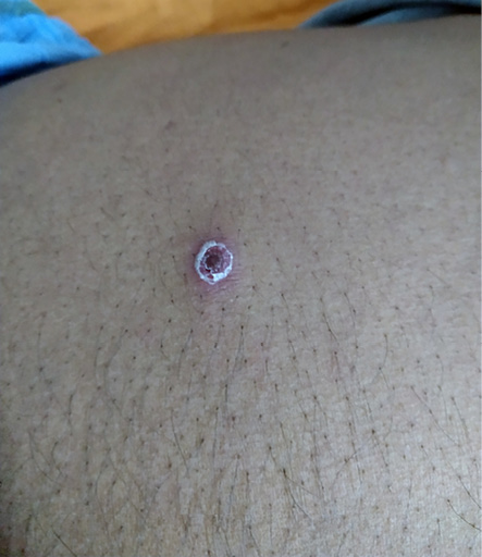 Figure 1: Clinical picture showing pathognomonic eschar on the abdomen.
Discussion
The onset of scrub typhus is characterized by fever, cough, headache, myalgia, and gastrointestinal symptoms.4 The characteristic findings include necrotic eschar, regional lymphadenopathy, interstitial pneumonia, encephalitis, and maculopapular rash. Studies have shown leukocytosis, transaminitis with or without cholestasis, and hepatosplenomegaly in patients with scrub typhus.5 The presenting spectrum of dengue fever is wide and includes high-grade fever, headache, retroorbital pain, transient blanching rash, and hemorrhagic complications, leading to rapid deterioration with development of shock syndrome.6 The generalized confluent erythematous rash with multiple small islets of normal skin often gives rise to the ‘white islands in red sea’ sign, a classic cutaneous manifestation of dengue.7 Isolation of dengue virus or detection by molecular methods such as reverse-transcription polymerase chain reaction is specific for diagnosing infection. However, in resource-poor countries like India, Nonstructural protein 1 (NS1) antigen assay along with serology (immunoglobulin M (IgM) and/or IgG) has been used successfully as a diagnostic tool.8 Recently, rapid diagnostic tests (RDTs) have been developed for the detection of dengue NS1 antigen and IgM/IgG antibodies, which are based mainly on immunochromatographic method. Results of such tests can be obtained within 30 minutes. Although RDT was previously considered a relatively inaccurate tool in diagnosis of dengue, a recently published study suggests that RDT had a high sensitivity and specificity in diagnosis, provided clinicians correlate its results with clinical signs and symptoms.9
Watt et al,10 made the following comparisons between dengue fever and scrub typhus. The median duration of fever at initial presentation is likely to be longer in scrub typhus (9 days) than in dengue (4 days).10 Lymphadenopathy is more common in scrub typhus (40%) than in dengue (22%). People having indoor occupations are more likely to present with dengue (51%) than with scrub typhus (15%).10 Bleeding manifestations such as bleeding from the gums tend to be more common in dengue fever than scrub typhus. The macular blanching rash in dengue is more subtle than in scrub typhus. A low leukocyte count (< 5000/mm3) and a low platelet count (< 140 000/mm3) are more commonly associated with dengue infections. Low platelet count is also a feature of scrub typhus but usually is associated with leukocytosis.10 Aspartate aminotransferase (AST) > Alanine aminotransferase (ALT) is found in both the diseases but in scrub typhus, elevated alkaline phosphatase is also seen.11 Severe thrombocytopenia, high AST and ALT with low albumin have been reported in coinfections.12
In our patient, the presence of thrombocytopenia with blanching rash favored dengue fever but the absence of hemorrhagic complications despite thrombocytopenia, elevated total leukocyte count, AST > ALT, and mildly elevated alkaline phosphatase favored a diagnosis of scrub typhus.
Reported cases of dengue and scrub typhus coinfection are rare in literature. One case which presented as acute febrile illness in a 50-year-old female from Nepal was reported by Sapkota et al.3 Another case of coinfection was reported from Uttar Pradesh (India) in a 45-year-old male with fever, hepatic and pancreatic dysfunction, and pneumonia, who responded well to oral doxycycline.13 Raina et al,13 evaluated dual infection as etiology of febrile illness in the sub-Himalayan region of north India, where they found dengue and scrub typhus coinfection in a few patients. Table 3 lists the previously published case reports and case series of dengue and scrub typhus coinfection.3,12–15
Therefore, a high degree of suspicion for coinfection should be made in cases of febrile illness in tropical countries, particularly in monsoon and post-monsoon seasons to prevent fatal consequences. Being recently traveling or living in hilly regions—the Himalayan foothills are common to most cases listed in Table 3—is also a factor to suspect dengue and scrub typhus coinfection.
Table 3: Comparison of clinical characteristics of current and previous reports on dengue and scrub typhus coinfection cases in the Indian subcontinent.
|
Sapkota et al3 (2017)
|
Southern belt, Nepal
|
50, Female
|
Regular visits to a nearby forest
|
Fever, headache, body ache, jaundice, lymphadenopathy. Eschar on right deltoid.
|
Leukocytosis, thrombocytopenia, elevated bilirubin, and transaminases.
|
Intravenous ceftriaxone and oral doxycycline for 7 days.
|
Discharged on 7th day.
|
|
Garg et al14 (2018)
|
Uttar Pradesh, India
|
45,
Male
|
Recent travel to sub-Himalayan region of Uttarakhand
|
High-grade fever, pedal edema, facial puffiness, and dyspnea.
Necrotic eschar on the dorsum of the penis.
|
Initial leukopenia followed by leukocytosis, thrombocytopenia, elevated bilirubin, transaminases, amylase, and lipase.
Bilateral bronchopneumonia, and features of pancreatitis on abdominal imaging.
|
Intravenous ceftriaxone and oral doxycycline for 7 days.
|
Discharged on 7th day.
|
|
Subedi et al15 (2021)
|
Western Nepal
|
33, Female
|
None
|
High fever, cough, dyspnea, and arthralgia followed by septic shock.
No eschar.
|
Leukocytosis, thrombocytopenia, transaminitis, renal impairment, and bilateral pulmonary infiltrates.
|
Oral doxycycline for 7 days.
|
Discharged on 7th day.
|
|
Basheer et al12 (2016)
Case series
|
South India
|
Mean age = 42.5,
males: 67%
|
Not mentioned
|
Fever in all, arthralgia in 3 patients, diffuse blanching rashes, and lymphadenopathy in 2 patients. Eschar noted in 2 patients (one in groin and other in axilla).
|
1 patient had leukocytosis, others had normal or reduced leukocytes count. Thrombocytopenia, hepatic transaminitis, and hypoalbuminemia were seen in
almost all.
|
Oral doxycycline for 4/6 patients. Azithromycin for 2/6 patients.
|
All responded to antibiotics within 48 hrs and became afebrile.
|
|
Raina et al13(2018)
Case series
|
Northern hilly state of Himachal Pradesh, India
|
Mean age = 36.6,
9/10 male
|
Out of 10 patients, 4 had no history of travel outside Himachal Pradesh
|
Fever in all, myalgia in 8, cough in 2, dyspnea, oliguria, seizure, and bleeding in 1 each.
|
8/10 patients had thrombocytopenia. Mean ALT = 114 IU/L and AST = 161 IU/L.
|
Not mentioned.
|
All the patients improved at the time of discharge.
|
ALT: alanine transaminase; AST: aspartate aminotransferase.
Conclusion
Due to overlapping clinical features and similar laboratory features, early diagnosis and treatment of cases of coinfection of dengue and scrub typhus are challenging to the physician. The coinfection possibility should be suspected early because delay in diagnosis can lead to longer hospital stay, increased chances of end-organ dysfunction, and mortality. A high index of suspicion should be made in patients from endemic areas presenting with febrile illness and multisystem involvement with deranged laboratory features and not responding to treatment. Meticulous knowledge of the physician regarding endemicity is the key in diagnosing such rare conditions.
Disclosure
The authors declared no conflicts of interest. Written consent was obtained from the patient.
references
- 1. Rapsang AG, Bhattacharyya P. Scrub typhus. Indian J Anaesth 2013 Mar;57(2):127-134.
- 2. Khetarpal N, Khanna I. Dengue fever: causes, complications, and vaccine strategies. J Immunol Res 2016;2016:6803098.
- 3. Sapkota S, Bhandari S, Sapkota S, Hamal R. Dengue and scrub typhus coinfection in a patient presenting with febrile illness. Case Rep Infect Dis 2017;2017:6214083.
- 4. Mahajan SK. Scrub typhus. J Assoc Physicians India 2005 Nov;53:954-958.
- 5. Hu ML, Liu JW, Wu KL, Lu SN, Chiou SS, Kuo CH, et al. Short report: abnormal liver function in scrub typhus. Am J Trop Med Hyg 2005 Oct;73(4):667-668.
- 6. Kamath SR, Ranjit S. Clinical features, complications and atypical manifestations of children with severe forms of dengue hemorrhagic fever in South India. Indian J Pediatr 2006 Oct;73(10):889-895.
- 7. Amrani A, Sil A, Das A. Cutaneous signs in infectious diseases. Indian J Dermatol Venereol Leprol 2021;88(4):569-575.
- 8. Muller DA, Depelsenaire AC, Young PR. Clinical and laboratory diagnosis of dengue virus infection. J Infect Dis 2017 Mar;215(suppl_2):S89-S95.
- 9. Yow KS, Aik J, Tan EY, Ng LC, Lai YL. Rapid diagnostic tests for the detection of recent dengue infections: an evaluation of six kits on clinical specimens. PLoS One 2021 Apr;16(4):e0249602.
- 10. Watt G, Jongsakul K, Chouriyagune C, Paris R. Differentiating dengue virus infection from scrub typhus in Thai adults with fever. Am J Trop Med Hyg 2003 May;68(5):536-538.
- 11. Thap LC, Supanaranond W, Treeprasertsuk S, Kitvatanachai S, Chinprasatsak S, Phonrat B. Septic shock secondary to scrub typhus: characteristics and complications. Southeast Asian J Trop Med Public Health 2002 Dec;33(4):780-786.
- 12. Basheer A, Iqbal N, Mookkappan S, Anitha P, Nair S, Kanungo R, et al. Clinical and laboratory characteristics of dengue-orientia tsutsugamushi co-infection from a tertiary care center in South India. Mediterr J Hematol Infect Dis 2016 Jun;8(1):e2016028.
- 13. Raina S, Raina RK, Agarwala N, Raina SK, Sharma R. Coinfections as an aetiology of acute undifferentiated febrile illness among adult patients in the sub-Himalayan region of north India. J Vector Borne Dis 2018 Apr-Jun;55(2):130-136.
- 14. Garg A, Jain A, Kashyap R. Travel-acquired scrub typhus infection masked by dengue fever in a patient from nonendemic area. J Glob Infect Dis 2018 Apr-Jun;10(2):114-115.
- 15. Subedi P, Ghimire M, Shrestha K, Ghimire K, Adhikari S, Tiwari B. Dengue and scrub typhus co-infection causing septic shock. Oxf Med Case Reports 2021 Dec;2021(11):omab115.