A 30-year-old Bangladeshi migrant worker with no underlying medical history, complained of multiple growths on his scrotum for the preceding three years [Figure 1]. It started with a single ‘pea-like’ swelling over the right scrotum which later grew in size and increased in numbers until both sides of the scrotum became affected. Aside from occasional itchiness, the growths were not associated with any ulceration, pain, or abnormal discharge. There was no history of preceding trauma, urogenital, or constitutional symptoms. Physical examination revealed multiple firm and nontender skin-colored nodules on both sides of the scrotum with the largest measuring 2 cm in diameter. There was no lymphadenopathy, and no similar lesion was found elsewhere on the body. Laboratory investigations reported normal levels of serum calcium, phosphorus, and parathormone. Excisional biopsy of the nodule showed multiple amorphous calcium deposits within the dermis free of epithelial lining or surrounding granulomatous reaction.

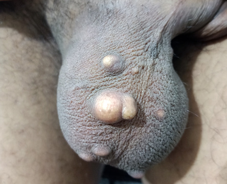 Figure 1: Multiple nodules on both sides of the scrotum.
Question
1. What is the diagnosis?
a. Scrotal calcinosis.
b. Nodular scabies.
c. Epidermal cyst.
d. Steatocystoma.
Answer
a. Scrotal calcinosis.
Discussion
Scrotal calcinosis is a rare condition marked by the presence of intradermal calcium deposits resulting in progressive appearance of skin nodules, which increase in number and size with age.1,2 Although benign, in extreme cases the disfigurement may be alarming. Scrotal calcinosis typically appears in early adulthood but patients tend to present late due to the indolent course of the disease.1,2 The disorder is mostly asymptomatic. Esthetic concern is the foremost reason for consultation.1 Occasionally, the lesions may be associated with itchiness, sense of pressure, chalky discharge, or ulceration.2,3
Current knowledge of this rare condition is limited to case reports and case series in the literature. As a result, the pathogenesis of the disease is uncertain. It was initially believed to be idiopathic, but emerging hypotheses suggest that the cause may be due to dystrophic calcification of pre-existent epidermal cyst, eccrine cyst, or degenerated dartos muscle.4,5 Foreign body infiltration and local trauma have been proposed as potential triggers for the calcification.2 None of the theories, however, has been conclusively proven.
The differential diagnosis for nodular scrotal growth include epidermal cyst, lymphangioma circumscriptum, steatocystoma, angiokeratoma, genital leiomyoma, epidermoid carcinoma, and nodular scabies among others.1,2,6 Nevertheless, Scrotal calcinosis is easily recognized clinically.2 Confirmation of diagnosis, however, is obtained through histopathology.4 Biopsy specimen stained with hematoxylin and eosin usually reveals the presence of calcium deposits within the dermis which can further be confirmed by applying the von Kossa stain. Epithelial lining is mostly absent with variable degree of surrounding granulomatous reaction.7
The mainstay of treatment is surgery which is indicated for intolerable local symptoms or esthetic improvement.2 Both single and multi-stage excision can be performed with good outcome although the lesions may recur.4 Excision is limited to the dermis leaving the dartos layer intact. Extensive disease may require partial scrotectomy with reconstruction.5 More recently, superior result was reported with the use of erbium YAG laser.2
Disclosure
The authors declared no conflicts of interest. Written informed consent was obtained from the patient for anonymized information to be published in this article.
references
- 1. Tareen A, Ibrahim RM. Idiopathic scrotal calcinosis - A case report. Int J Surg Case Rep 2018;44:51-53.
- 2. Fernández-Nieto D, de Perosanz-Lobo D, Jiménez-Cauhé J, Ortega-Quijano D, Jaén-Olasolo P. A middle-aged man with indurated scrotal nodules. Aust J Gen Pract 2019 Jul;48(7):463-464.
- 3. Kaur G, Phogat D, Trehan A. Idiopathic scrotal calcinosis.
J Mar Med Soc 2017;19(2):131-133.
- 4. Akinboro AO, Onilede DA, Babatunde TO, Oiwoh SO, Suleiman OA, Olabode OP. Idiopathic scrotal calcinosis: report of 2 cases, and review of pathogenesis and factors that determine patients’ acceptance of surgical treatment. Clin Cosmet Investig Dermatol 2018 Jul;11:333-337.
- 5. Jayarajah U, de Silva L, de Silva C, Seneviratne S. Idiopathic scrotal calcinosis: a case report of a rare entity. Case Rep Urol 2019 Dec;2019:6501964.
- 6. Wollina U, Schönlebe J, França K, Tchernev G, Lotti T. Idiopathic scrotal calcinosis – a case report. Open Access Maced J Med Sci 2018 Jan;6(1):108-109.
- 7. Kilitci A, Kaya Z, Acar EM, Elmas ÖF. Scrotal calcinosis: analysis of 5 cases. J Clin Exp Investig 2018;9(4):150-153.