A two-month-old female presented to the local emergency department with respiratory distress in the setting of a three-day history of febrile illness with symptoms of an upper respiratory infection. During initial evaluation, she was noted to be lethargic, tachycardic, and tachypneic. She was given fluid resuscitation, and a sepsis evaluation was initiated. However, soon afterward she developed acute hypoxemic respiratory failure with oxygen saturations in the mid to low 80s that were refractory to non-invasive positive pressure ventilation. She subsequently went into cardiac arrest and required five minutes of cardiopulmonary resuscitation before the return of spontaneous circulation. She was intubated during the resuscitative phase and placed on invasive mechanical ventilatory support. Despite conventional therapy for acute respiratory failure, the patient continued to be hypoxemic, and breath sounds were noted to be asymmetrical. Thus, a chest radiograph was obtained [Figure 1]. After stabilization, the patient was referred to a tertiary medical center for further care.
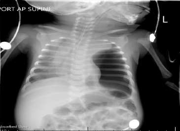
Figure 1: Chest radiograph of a two-month-old female with a large air collection in the left hemithorax with a thin edge identified at the superior left lateral aspect and a rightward shift of the heart and mediastinum.
Question
- What is the likely diagnosis?
a. Acute pneumothorax.
b. Congenital diaphragmatic hernia.
c. Endotracheal tube malposition.
d. Acute bacterial pneumonia.
Answer
b. Congenital diaphragmatic hernia.
Discussion
Congenital diaphragmatic hernia (CDH) is an anatomical birth defect in which the diaphragm incompletely forms, resulting in abdominal viscera protruding into the thoracic cavity.1 It is estimated to occur with a frequency of approximately 1 to 5 cases in 10 000 live births worldwide.1 As a direct result of abdominal viscera herniating into the thoracic cavity, organ development within the thoracic cavity is often affected. This can lead to pulmonary hypoplasia, alterations in pulmonary vasculature, and left ventricular hypoplasia.2 The severity of clinical presentation is related to the degree to which the structures within the thoracic cavity are altered; thus, neonates are at risk for hypoxemia, pulmonary hypertension, and hemodynamic instability.3 The mortality risk of this condition is high when it presents at birth.4
Five to twenty-five percent of CDH cases can present after the neonatal period with respiratory and/or gastrointestinal symptoms.5,6 Treatment depends on the clinical circumstances. If found incidentally, the patient can be observed until definitive surgical correction is performed.7 In patients with respiratory failure, treatment is provided with gentle, supportive respiratory support (while carefully avoiding further lung injury) and gastric decompression via nasogastric tube.8 After stabilization or if the patient has acute abdominal symptoms, the patient is referred for surgical intervention.7 Late presenting CDH patients tend to have a better long-term prognosis as they do not typically exhibit pulmonary hypoplasia or pulmonary hypertension.9
When a pediatric patient in acute respiratory failure remains hypoxemic despite standard treatments, further evaluation is required to determine the etiology. Common conditions that should be considered include endotracheal tube dislodgement, airway obstruction, equipment failure, and acute pneumothorax. Late-onset CDH, while not a common condition resulting in hypoxemia, can also present with symptoms of acute respiratory distress. Specifically, it can mimic a tension pneumothorax both clinically and radiographically in the setting of gastric distension after non-invasive positive pressure ventilation.8 Therefore, early recognition and a high index of suspicion are necessary to prevent iatrogenic complications associated with misdiagnosis.
Disclosure
The authors declared no conflicts of interest.
references
- 1. Chandrasekharan PK, Rawat M, Madappa R, Rothstein DH, Lakshminrusimha S. Congenital diaphragmatic hernia - a review. Matern Health Neonatol Perinatol 2017 Mar;3(1):6.
- 2. Patel N, Massolo AC, Kipfmueller F. Congenital diaphragmatic hernia-associated cardiac dysfunction. Semin Perinatol 2020 Feb;44(1):151168.
- 3. Gupta VS, Harting MT. Congenital diaphragmatic hernia-associated pulmonary hypertension. Semin Perinatol 2020 Feb;44(1):151167.
- 4. Harting MT, Lally KP. The congenital diaphragmatic hernia study group registry update. Semin Fetal Neonatal Med 2014 Dec;19(6):370-375.
- 5. Bagłaj M. Late-presenting congenital diaphragmatic hernia in children: a clinical spectrum. Pediatr Surg Int 2004 Sep;20(9):658-669.
- 6. Kitano Y, Lally KP, Lally PA; Congenital Diaphragmatic Hernia Study Group. Late-presenting congenital diaphragmatic hernia. J Pediatr Surg 2005 Dec;40(12):1839-1843.
- 7. Chao P-H, Chuang J-H, Lee S-Y, Huang H-C. Late-presenting congenital diaphragmatic hernia in childhood. Acta Paediatr 2011 Mar;100(3):425-428.
- 8. Kajal P, Bhutani N, Goyal M, Kamboj P. Iatrogenic gastric perforation in a misdiagnosed case of late presenting congenital diaphragmatic hernia: report of an avoidable complication. Int J Surg Case Rep 2017;41:154-157.
- 9. Akinkuotu AC, Cruz SM, Abbas PI, Lee TC, Welty SE, Olutoye OO, et al. Risk-stratification of severity for infants with CDH: Prenatal versus postnatal predictors of outcome. J Pediatr Surg 2016 Jan;51(1):44-48.