Uncommon Presentation of Congenital Lacrimal Duct Obstruction A Case Report of Congenital Amniontocele
Jagdish Bhatia, Najmi Rahman, Mathew Varghese
ABSTRACT
Background: Congenital amniontocele also known as congenital dacryocystocele is an uncommon variant of a very common condition (Naso-Lacrimal Duct Obstruction, NLDO) that affects newborn babies. The congenital amniontocele causes obstruction of the lacrimal drainage system creating a cystic swelling of the lacrimal sac. It usually presents at birth or soon after birth, as a bluish swelling, below and nasal to the medial canthus. If the congenital amniontocele is associated with intranasal cystic swelling of the NLD and if it is large and bilateral it can cause severe respiratory distress particularly during milk feeding. Method: We report a case of 24 days old female newborn baby who were presented to us from pediatric clinic of Rustaq Hospital with bluish cystic swelling below the left medial canthus soon after the birth. Conclusion: Congenital amniontocele is relatively rare and the literature describes a variable natural course of these lesions with resultant controversy in their management. Usually congenital amniontocele resolves spontaneously with conservative management, and if not, dacryocystitis might develop. Treatment of congenital amniontocele comprises digital sac massaging and early probing the lacrimal system,8 or some times, intranasal surgery in the form of marsupialization of the intranasal cyst, this is usually performed endoscopically.7 We report successful conservative management in our case and recommend this with oral and topical antibiotics with lacrimal sac massage for 7 to 10 days and, if there is no response, to consider surgical intervention in the form of lacrimal probing.
Keywords: Congenital amniontocele, dacryocystocele, lacrimal probing, antibiotics, conservative.
Submitted: 15 November 2006
Revieved: 29 August 2007
Accepted: 12 September 2007
From the Department of Ophthalmology, Rustaq Hospital, From the Rustaq, Sultanate of Oman
Address correspondence and reprint request to: Jadish Bhatia, Department of Ophthalmology, Rustaq Hospital, P.O. Box 421, P.C. 329, Rastaq, Sultanate of Oman. E-Mail: imbhatia@gmail.com
INTRODUCTION
Infants rarely present with tense visible swelling of the lacrimal sac, a congenital amniontocele, also known as congenital dacryocystocele which occurs when there is a combination of congenital lacrimal obstruction and obstruction to retrograde flow through the canaliculi. There is a strong female preponderance (73%) and unilateral involvement (88%).1 The origin and composition of the sac contents have been discussed for years in the ophthalmic literature. Most believe that the fluid in the amniontocele is amniotic fluid, since the tear experts secretion normally is not significant until 3 to 4 weeks postpartum. However, because of the viscous nature of the sac contents, some authors have proposed that it represent mucus produced by the intraluminal goblet cells.2
This swelling usually has a definite blue discoloration, often misdiagnosed as a vascular lesion. The infant encounters no clinical signs of epiphora (tearing) or dacryocystitis (infection). The amniontocele is typically sterile since colonization of the lacrimal drainage system is usually not established until the first several weeks of life. When infected, the most common bacteria isolated are Staphylococcus organisms.3
The clinical course is distinct from that of uncomplicated very common congenital naso-lacrimal duct obstruction: Acute inflammation is common, developing in and around the lacrimal sac within a few days of birth in 20-60% of patients.1, 3 In a minority of cases there is communication down the nasolacrimal duct due to a large intranasal cyst; this is a well documented cause of neonatal respiratory obstruction, which may require urgent treatment.1, 4
CASE REPORT
A 24 days old female newborn baby presented to us from pediatric clinic of Rustaq Hospital with the history of swelling below the left medial canthus, soon after birth. On examination, there was a bluish colored swelling below the left medial canthus.
(Figure 1) Otherwise anterior segment of both the eyes were normal. Compression of lacrimal sac area did not show any regurgitation of fluid. There was no history of watering or discharge from the affected eye. Pediatric consultation was unremarkable. Baby was born full term with normal antenatal history.
After clinical examination, a diagnosis of congenital amniontocele was made and child was put on oral antibiotics (Amoxicillin oral drops) and topical antibiotic drops (Gentamicin eye drops) with instructions of sac massage. She was advised follow up after one week. She reported back to us after one week with complete resolution of sac swelling. (Figure 2) Oral and topical antibiotic were continued for one more week. During next follow up after 1 week, amniontocele completely regressed and the child was symptom free. The patient was discharged from the eye clinic with the advice of regular follow up in eye clinic for monitoring.
DISCUSSION
There are few studies to guide management. While some reports have advised immediate probing of the lacrimal passages, most recommend a delay of 5-7 days, as spontaneous resolution may occur with conservative management.1, 3 If signs of inflammation develop, immediate probing should be performed and treatment with systemic antibiotics started.5 Those in favor of early probing claim that by relieving the obstruction, atony of the sac and disruption of the medial canthal musculature and skin are prevented, preserving the lacrimal pump mechanism. Furthermore, if left untreated, the fluid filled sac is at a higher risk of infection, secondary scarring and permanent closure, requiring a more involved surgical by-pass procedure termed dacryocysto-rhinostomy.6

Figure 1: Congenital amniontocele. Soon after birth.
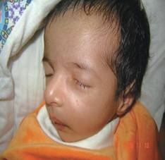
Figure 2: One week after conservative management.
Complete resolution
CONCLUSION
True congenital amniontocele is a relative rarity. The current case demonstrates successful management of congenital amniontocele with early conservative management with oral and topical antibiotics and digital sac massage. Surgical intervention is indicated in cases of cellulites, breathing difficulty from large nasal cyst, recurrent amniontocele, and lack of its resolution after a short trial of conservative management for 1 to 2 weeks. We recommend conservative management with oral and topical antibiotics with lacrimal sac massage initially for 7 to 10 days and, if there is no response then to consider surgical intervention in the form of lacrimal probing.
REFERENCES
-
Mansour AM, Cheng KP, Mumma JV, Stager DR, Harris GJ, Patrinely JR, et al. Congenital dacryocele. A collaborative review. Ophthalmology 1991; 98:1744-1750.
-
Calhoun JH. Problems of lacrimal system in children. Pediatr Clin North Am. 1987; 34:1457-1465.
-
Weinstein GS, Biglan AW, Patterson JH. Congenital lacrimal sac mucoceles Am J Ophthalmol 1982; 94:106-110.
-
Grin TR, Mertz JS, Stass-Isern M. Congenital nasolacrimal duct cysts dacryocystocele. Ophthalmology 1991; 98:1238-1242.
-
Pollard ZF. Treatment of acute dacryocystitis in neonates. J Pediatr Ophthalmol Strabismus 1991; 28:341-343.
-
Andrea Tardiff, Harvey P, Cole III, Ralph E, et al. Dacryocele. www.Fetus.net
-
Hepler KM, Woodson GE, Kearns DB. Respiratory distress in the neonate Sequela of a congenital dacryocystocele. Arch Otolaryngol Head Neck Surg. 1995; 121:1423-1425.
-
Steinkogler FJ. Primary lacrimal duct probing in congenital dacryocystitis Disadvantages and alternatives. Padiatr Padol. 1985; 20:185-191.