Bullous systemic lupus erythematosus (BSLE) is an autoimmune and chronic bullous dermatosis. It is characterized by distinctive clinical and histological features that can occur in the course of systemic lupus erythematosus (SLE) or its initial presentation.1,2 BSLE usually manifests in the second and third decades of life, most frequently in black women.3 Less than 5% of patients with SLE develop vesiculobullous lesions in isolation or along with other cutaneous manifestations.4 However, only a few cases have been reported in children.5 We describe the case of a young girl with recalcitrant SLE and nephritis who initially presented with widespread vesiculobullous lesions and fulfilled the criteria of BSLE.
case report
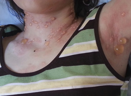
Figure 1: Vesiculobullous eruptions on the patient’s skin.
A 14-year-old girl initially presented to our institution with low-grade fever and fatigue. One month later, she developed widespread vesiculobullous skin lesions, accompanied with erosions and crusting. On admission, she was febrile and had necrotic ulcers over the hard palate and oral mucosa [Figure 1], and tense grouped vesicle and bulla on her face, neck, trunk, extremities, and genital area. Nikolsky's sign was negative, with no malar or discoid rash, generalized lymphadenopathy, or hepatosplenomegaly. Respiratory and cardiovascular examinations were normal. She had no history of photosensitivity.
Blood investigations revealed a hemoglobin concentration of 6.9 g/dL, white blood cells of 7 × 10⁹/L, and neutrophils of 4.9 × 10⁹/L. Her lymphocytes level was 2 × 10⁹/L, and platelets were 653 × 109/L. Her erythrocyte sedimentation rate (ESR) was 120 mm at the first hour, and C-reactive protein (CRP) was negative. Her serum creatinine level was 0.8 mg/dL, and blood urea nitrogen (BUN) was 34 mg/dL. A peripheral blood smear showed anisocytosis, and target cell and reticulocyte was 3.5% (normal range: 1−1.5%). Direct and indirect Coombs tests were negative. Serologic tests to rule out SLE and other collagen vascular diseases showed antinuclear antibody (ANA) levels of 180 AU/mL (> 25 positive) and anti-double-stranded DNA (anti-dsDNA) of 260 IU/mL (> 100 positive). Anticardiolipin immunoglobulin (Ig)-G and IgM antibodies, lupus anticoagulant, anti-Ro, anti-La, anti-Sm antibodies, and the venereal disease research laboratory (VDRL) test were negative.
Complement components assay revealed C3 levels of 0.04 gr/L (normal range: 0.89−1.7), C4 of 0.03 gr/L (normal range: 0.16−0.3), 14% CH50 (normal range: 76−220), and C1q of 5.3 µg/mL
(> 15 positive). Urine microscopy showed hematuria and proteinuria. Human immunodeficiency virus (HIV)-1, HIV2, hepatitis B surface antibody, and hepatitis C virus (HCV) antibodies were negative. Lesion swabs were negative for herpes simplex virus antigen.
Histological examination of the lesion showed separation of the dermis, epidermis, and subepidermal bulla. Direct immunofluorescence (DIF) demonstrated a strong linear deposition of IgG and less IgA and IgM, and also complements’ (C3) deposition at the dermoepidermal junction.
The renal biopsy indicated diffuse glomerular basement membrane thickening and marked spike patterns in silver stain. Immunofluorescence studies revealed granular deposition of IgG, IgA, IgM, C3, and C1q in the capillary wall of the glomerulus, indicative of membranous nephropathy, suggestive of class V lupus nephritis with no chronicity. Bone marrow aspiration detected myeloid hyperplasia.
Based on the clinical investigations, the patient was diagnosed with BSLE with lupus nephritis. The case fulfilled the criteria of BSLE.6 She received three methylprednisolone pulses (30 mg/kg/day) and hydroxychloroquine (5 mg/kg) for three subsequent days. However, it did not alleviate the vesiculobullous eruption and dapsone (50 mg/day) was given, resulting in a dramatic disappearance of the lesions. Dapsone interruption due to hemolysis did not aggravate the bullous disease. Meanwhile, hemolysis and a drop in hemoglobin levels continued, so intravenous immunoglobulin (IVIG) was started (1 g/kg) for two days, after which the fever and hemolysis stopped. The patient was discharged with oral prednisolone and hydroxychloroquine.
On follow-up, the patient had recurrent flare-ups of disease and aggravation of renal disease. Despite aggressive treatment, ESR and anti-dsDNA levels did not decrease. We added cyclophosphamide pulse to the previous treatment according to protocols for lupus nephritis. The patient's anti-dsDNA levels returned to normal (15 IU/mL). Proteinuria decreased eight months after initial presentation, although nephritis was not completely controlled. The patient continues to be monitored as an outpatient, and her skin bullous lesions have not returned.
discussion
Blisters in SLE can be because of BSLE or SLE with blisters. Subepidermal blister formation in the course of severe SLE occurs due to extensive interface inflammation and basal cell vacuolation, showing polycyclic erosions with an advancing blistering border predominantly on sun-exposed areas. But BSLE, histologically, is a subepidermal blistering disease with an acute neutrophil-predominant infiltrate in the upper dermis.7,8
In this report, we described the case of a young girl with BSLE as an initial manifestation. The diagnosis of BSLE type 1 requires five elements: 1. Fulfillment of the American College of Rheumatology criteria for SLE; 2. Acquired vesiculobullous eruption; 3. Histological evidence of a subepidermal blister and a predominantly neutrophilic dermal infiltrate; 4. DIF microscopy demonstrating IgG and IgM deposits at the basement membrane zone; and 5. Evidence of antibodies to type VII collagen by DIF or indirect immune fluorescence on salt-split skin, immune blotting, immune precipitation, enzyme-linked immune sorbent assay, or immune electron microscopy.6
Type 2 BSLE is characterized by the presence of first four criteria although the location of the antigen is undefined or the dermal antigen is other than type VII collagen. In type 3 BSLE the location of the antigen is epidermal.9,10
Our patient fulfilled all BSLE criteria although autoantibodies could not be demonstrated to type VII collagen to determine the type.
Type VII collagen is the major component of anchoring fibrils at the dermal-epidermal junction and the main target of immune system in BSLE such as epidermolysis bullosa acquisita. However, a recent study of BSLE patients demonstrated that autoantibodies to other elements of the basement membrane including laminin-5, laminin-6, and bullous pemphigoid (BP) antigen I are effective in the pathogenesis of BSLE.1,11
The relationship between bullous eruptions and symptom flares in SLE has not been determined. Some studies found that vesiculobullous lesions can develop without clinical or laboratory evidence of disease worsening.2,12 In our case, bullous eruptions disappeared with dapsone and, despite multiple flare-ups of systemic disease, did not return.
The pathogenesis of developing nephritis in BSLE is not clear. Type VII collagen is usually not present in normal glomeruli. It seems that glomerular and/or tubular scarring, regardless of the underlying disease, results in injury to renal extracellular matrix and de novo expression of fibrillary collagens. Hence, this newly performed epitope can trigger autoimmunity reactions.13,14
Our case had membranous glomerulonephritis (class V lupus nephritis), which was progressive and resistant to different immunosuppressive treatments. There was no association between kidney disease activity and bullous lesions. However, in some cases, a clear association between skin manifestations and renal activity is seen. Our case illustrates that a generalized vesiculobullous lesion can be the initial presentation of SLE.
Conclusion
Differentiation between BSLE and SLE with blister is important because BSLE may be a risk for developing lupus nephritis and may relate to worse prognosis and refractory disease. The unusual presentation of this disease may delay the diagnosis and therefore requires a high index of clinical suspicion.
Disclosure
The authors declared no conflicts of interest.
references
- Duan L, Chen L, Zhong S, Wang Y, Huang Y, He Y, et al. Treatment of Bullous Systemic Lupus Erythematosus. Journal of Immunology Research. 2015;2015:167064.
- 2. Fujimoto W, Hamada T, Yamada J, Matsuura H, Iwatsuki K. Bullous Systemic Lupus Erythematosus as an Initial Manifestation of SLE. J Dermatol 2005 Dec;32(12):1021-1027.
- 3. Miziara ID, Mahmoud A, Chagury AA, Alves RD. Bullous Systemic Lupus Erythematosus: Case report. Int Arch Otorhinolaryngol 2013 Jul;17(3):344-346.
- 4. Dhir R, Desylva P, Gehi N, Malik A, Singh YD, Jagannayakulu H, et al. Pericardial effusion with vesiculobullous lesions in a young female. Bullous systemic lupus erythematosus (bullous SLE). Indian J Dermatol Venereol Leprol 2006 Mar-Apr;72(2):175-177.
- 5. Lourenço DM, Gomes RC, Aikawa NE, Campos LM, Romiti R, Silva CA. Childhood-onset bullous systemic lupus erythematosus. Lupus 2014 Nov;23(13):1422-1425.
- 6. Camisa C, Grimwood RE. Indirect immunofluorescence in vesiculobullous eruption of systemic lupus erythematosus. J Invest Dermatol 1986 May;86(5):606.
- 7. Burrows NP, Bhogal BS, Black MM, Rustin MH, Ishida-Yamamoto A, Kirtschig G, et al. Bullous eruption of systemic lupus erythematosus: a clinicopathological study of four cases. Br J Dermatol 1993 Mar;128(3):332-338.
- 8. Yung A, Oakley A. Bullous systemic lupus erythematosus. Australas J Dermatol 2000 Nov;41(4):234-237.
- 9. Yell JA, Allen J, Wojnarowska F, Kirtschig G, Burge SM. Bullous systemic lupus erythematosus: revised criteria for diagnosis. Br J Dermatol 1995 Jun;132(6):921-928.
- 10. Patrício P, Ferreira C, Gomes MM, Filipe P. Autoimmune bullous dermatoses: a review. Ann N Y Acad Sci 2009 Sep;1173:203-210.
- 11. Chan LS, Lapiere JC, Chen M, Traczyk T, Mancini AJ, Paller AS, et al. Bullous systemic lupus erythematosus with autoantibodies recognizing multiple skin basement membrane components, bullous pemphigoid antigen 1, laminin-5, laminin-6, and type VII collagen. Arch Dermatol 1999 May;135(5):569-573.
- 12. Ng YY, Chang IT, Chen TW, Liou HN, Yang AH, Yang WC. Concomitant lupus nephritis and bullous eruption in systemic lupus erythematosus. Nephrol Dial Transplant 1999 Jul;14(7):1739-1743.
- 13. Onetti Muda A, Ruzzi L, Bernardini S, Teti A, Faraggiana T. Collagen VII expression in glomerular sclerosis. J Pathol 2001 Oct;195(3):383-390.
- 14. Sáez-de-Ocariz M, Espinosa-Rosales F, López-Corella E, de León-Bojorge B; Sáez-de-OcarizM. Bullous lesions as a manifestation of systemic lupus erythematosus in two Mexican teenagers. Pediatr Rheumatol Online J 2010;8:19.