Sternoclavicular joint dislocation (SCJD) is a rare entity, accounting for less than 1% of all dislocations in the body and approximately 3% of all shoulder girdle injuries. Of all the SCJD cases, 90% to 95% are anterior, and the rest are posterior. These types of injuries are mainly associated with high-impact trauma, but they can also occur as sports injuries or sometimes simple falls.1,2 Posterior SCJD can be life-threatening because of its relation to mediastinal structures. It can cause tracheal compression, injury to the brachial plexus, vocal cord paralysis, pneumothorax, respiratory compromise, and vascular injuries.3 Most of the complications are acute, but late-appearing complications are observed in unreduced posterior dislocations and include thoracic outlet syndrome, brachial neuropathy, subclavian artery stenosis, exertional dyspnea, and septicemia following tracheoesophageal fistula. The main treatment of anterior SCJD is closed reduction. For posterior SCJD, urgent closed reduction is advised, and if it fails, operative management is warranted.4 Various surgical techniques have been used, such as osteosynthesis with pins and K wires, plate fixation, and ligament fixation. We describe a figure-of-eight reconstruction technique using the semitendinosus tendon as an autograft in a patient with traumatic posterior SCJD.
Case Report
A 21-year-old male patient was admitted to Nizwa Hospital following a high-impact motor vehicle accident. On arrival, the patient was conscious and oriented with a Glasgow Coma Scale score of 15. He complained of left-sided chest pain with tenderness at the left sternoclavicular joint (SCJ), and severe pain with movement of the left arm and shoulder. The chest X-ray showed a posterior left SCJ dislocation [Figure 1]. Computed tomography (CT) scan of the chest with 3D reconstruction confirmed the posterior dislocation of the left SCJ, with displacement of the trachea to the right along with compression of the mediastinal structure’s posterior to the joint [Figure 2].
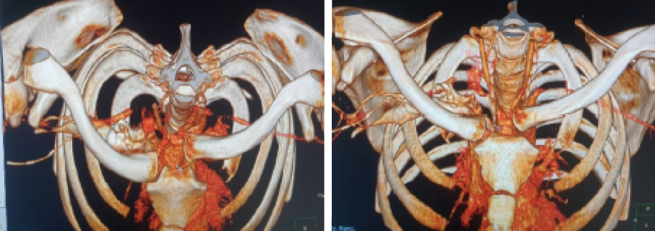 Figure 1: Preoperative computed tomography reconstructed 3D image showing left posterior dislocation of the left sternoclavicular joint.
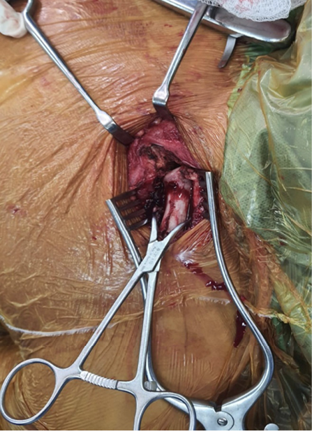 Figure 2: Operative image.
Closed reduction was attempted but was not successful. It was decided to resort to open reduction with fixation and reconstruction of the joint. The patient was positioned in the beach chair position, and the shoulder, entire clavicle, and sternum were exposed. The arm was placed by his side, and the contralateral leg was prepared to harvest the semitendinosus tendon. The semitendinosus autograft was harvested first using the standard technique. A 4 cm incision was made at the point of insertion of the pes anserinus. The sartorius fascia was incised, exposing the semitendinosus tendon on its undersurface. Approximately 16 cm of the tendon was harvested. The graft was cleared of all the tissue. The diameter of the tendon was required to be such that it could pass through a 4.8 mm drilled tunnel. Number 2 Ethibond™ suture was used as a locking whipstitch at the free end of the tendon to help in passing it through the tunnel. The graft was kept in saline-soaked gauze. An 8 cm incision was made over the left SCJ. The medial end of the clavicle and manubrium was exposed subperiosteally using a periosteal elevator, taking care not to injure the underlying neurovascular structures. To fully reduce the SCJ, a small portion of the medial part of the clavicle was excised. After clearing the hematoma, the joint was identified, and the intra-articular disc was removed.
Once satisfied with the reduction, the bone was drilled to create a tunnel for the passage of the tendon. Underlying structures were protected by placing a spatula beneath while drilling the bone. The first two parallel holes were drilled along the articulating edge of the clavicle, then similar holes were drilled along the articulating edge of the manubrium. The edges of the holes were smoothed with a small curette to ease the passing of the tendon. Shuttling Vicryl® sutures No. 0 were placed in the tunnels for threading the graft into the tunnel. The graft was passed in a figure-of-eight configuration, with the limbs of the graft crossing over the joint anteriorly. Care was taken so that the graft slid freely in the tunnels. The SCJ was reduced by putting tension on the traction sutures at the end of the two limbs of the graft. While maintaining the reduction, multiple 2-0 Ethibond sutures were placed on different limbs of the graft and on the knot to secure the graft from loosening. After ensuring the stability of the joint and graft tension, the wound was closed in layers. Postoperative CT reconstruction showed good and stable repair, with a reduced joint [Figure 3].
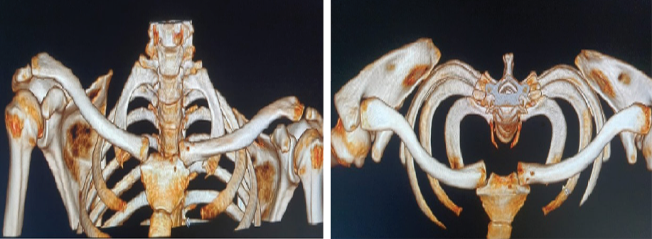 Figure 3: Postoperative reconstructed CT image showing reduced joint after figure-of-8 reconstruction technique using semitendinosus tendon.
Postoperatively, the patient’s arm was immobilized by a sling for four weeks. Thereafter, physiotherapy involving assisted motion was initiated with an initial maximum abduction to 90 degrees. The range of motion and weight-bearing were progressively increased according to the clinical course. Informed consent was obtained from the patient.
Discussion
The dislocation of the SCJ is rare.5 Anterior SCJD cases occur three times as often as posterior dislocation cases, due to the relative weakness of the anterior sternoclavicular ligament. The leading cause of posterior SCJD is high-energy trauma following a direct blow to the posterolateral part of the shoulder with the arm in flexion and adduction.6 Many of these dislocations are attributed to sports injuries, but they are also seen after traffic accidents, as in our patient, or falls.7
The SCJ is important in articulation for the movement of the shoulder girdle and is the only bony attachment of the arm to the axial skeleton. It is a saddle-type synovial joint between the medial end of the clavicle, the clavicular notch of the manubrium, and the upper medial surface of the first costal cartilage. The joint is stabilized by the anterior sternoclavicular ligament anteriorly and the stronger posterior sternoclavicular ligament posteriorly. The continuation of the deep cervical fascia forms the interclavicular ligament and connects the medial heads of the two clavicles. The costoclavicular ligament connects the clavicle to the first rib and the first costal cartilage. An injury to all these ligaments will cause the posterior dislocation of the joint.8
The usual presentation is pain around the medial aspect of the clavicle with shoulder and neck movement, along with a gap in the medial aspect of the clavicle with swelling. Other symptoms depend on injury to adjacent structures, like dyspnea, hoarseness of voice, or dysphagia. If the injury is initially missed, it may present later with tracheal stenosis, thoracic outlet syndrome, vascular injury, or brachial plexopathy.6,7
X-ray imaging with standard views might miss the injury, is often inaccurate, and is hard to interpret. Special views like the serendipity view and 40-degree cephalic tilt may provide some insight. CT scan or magnetic resonance imaging scan is ideal for diagnosing the injury. They also help to assess any damage to the surrounding structures, particularly those lying posterior to the SCJ. A contrast study is required to rule out any vascular injury.
While anterior SJCD can usually be managed conservatively, posterior SJCD requires a more aggressive approach. The preferred method of treatment is closed reduction, preferably in the operating theater because of the proximity of intrathoracic and mediastinal structures. Tepolt et al,9 in their meta-analysis, found that the chances of success in closed reduction were low if performed later than 48 hours. If closed reduction fails, then open reduction with internal fixation is advised, with a cardiothoracic surgeon on standby. A cardiothoracic surgeon is required in case there is damage to the vessels behind the SCJ in posterior dislocation, or to deal with any injury to mediastinal structures while mobilizing the joint or drilling the bone.
There is a lack of consensus on the optimal operative technique for posterior SJCD because of its rarity. Various techniques have been described to achieve a stable sternoclavicular joint through open reduction internal fixation surgeries. These include ligament repair with reconstruction,10 plate fixation,11 and figure-of-eight reconstruction using various tendons. According to the literature, ligament repair with reconstruction is the most used method.12 Its advantages include superior biomechanical stability, particularly in the posterior direction. The use of autologous tissue improves healing, reduces the risk of infection, and enhances tissue integration.13 Kitchener wire and pin are now contraindicated because of the reported cases of their migration into vital structures.14
Conclusion
Posterior SCJD cases are extremely rare, but due to the risk of injuring the innominate vein and subclavian vessels, prompt action is required. If a stable closed reduction is not possible, then open reduction with fixation is necessitated. Several techniques for SCJ fixation have been described, but there is no consensus on which technique is better because of the rarity of the injury. In the present case, we used the figure-of-eight reconstruction technique using the semitendinosus tendon with satisfactory results.
Disclosure
The authors declare no conflicts of interest.
references
- 1. Ngom G, Mohamed AS, El Housseine MO, Ndour O. Traumatic posterior sternoclavicular joint dislocation in a child: a case report. Pan Afr Med J 2014 Dec;19:386.
- 2. Bensafi H, Laffosse J-M, Taam SA, Molinier F, Chaminade B, Puget J. Tamponade following sternoclavicular dislocation surgical fixation. Orthop Traumatol Surg Res 2010 May;96(3):314-318.
- 3. Worman LW, Leagus C. Intrathoracic injury following retrosternal dislocation of the clavicle. J Trauma 1967 May;7(3):416-423.
- 4. Robinson CM, Jenkins PJ, Markham PE, Beggs I. Disorders of the sternoclavicular joint. J Bone Joint Surg Br 2008 Jun;90(6):685-696.
- 5. Elting JJ. Retrosternal dislocation of the clavicle. Arch Surg 1972 Jan;104(1):35-37.
- 6. Cope R. Dislocations of the sternoclavicular joint. Skeletal Radiol 1993;22(4):233-238.
- 7. Garg S, Alshameeri ZA, Wallace WA. Posterior sternoclavicular joint dislocation in a child: a case report with review of literature. J Shoulder Elbow Surg 2012 Mar;21(3):e11-e16.
- 8. Rajaratnam S, Kerins M, Apthorp L. Posterior dislocation of the sternoclavicular joint: a case report and review of the clinical anatomy of the region. Clin Anat 2002 Mar;15(2):108-111.
- 9. Tepolt F, Carry PM, Heyn PC, Miller NH. Posterior sternoclavicular joint injuries in the adolescent population: a meta-analysis. Am J Sports Med 2014 Oct;42(10):2517-2524.
- 10. Armstrong AL, Dias JJ. Reconstruction for instability of the sternoclavicular joint using the tendon of the sternocleidomastoid muscle. J Bone Joint Surg Br 2008 May;90(5):610-613.
- 11. Shuler FD, Pappas N. Treatment of posterior sternoclavicular dislocation with locking plate osteosynthesis. Orthopedics 2008 Mar;31(3):273.
- 12. Spencer EE, Kuhn JE, Huston LJ, Carpenter JE, Hughes RE. Ligamentous restraints to anterior and posterior translation of the sternoclavicular joint. J Shoulder Elbow Surg 2002;11(1):43-47.
- 13. Wang D, Camp CL, Werner BC, Dines JS, Altchek DW. Figure-of-8 reconstruction technique for chronic posterior sternoclavicular joint dislocation. Arthrosc Tech 2017 Oct;6(5):e1749-e1753.
- 14. Lyons FA, Rockwood CA Jr. Migration of pins used in operations on the shoulder. J Bone Joint Surg Am 1990 Sep;72(8):1262-1267.