Cornual ectopic pregnancy is a rare and potentially life-threatening type of ectopic pregnancy. The diagnosis of cornual ectopic pregnancy is intricate, and the condition necessitates swift intervention. Laparoscopic methods, such as resection and salpingectomy, are effective surgical choices that can minimize blood loss and mitigate the likelihood of future complications.1 We present the case of a 37-year-old multiparous woman diagnosed with a cornual ectopic pregnancy who was successfully managed.
Case Report
A 37-year-old woman (gravida 6, para 3), presented to the emergency department with right-sided lower abdominal pain and vaginal spotting at six weeks and two days of gestation. She had a history of three uncomplicated cesarean section deliveries and one cornual ectopic pregnancy that was managed by laparotomy myomectomy and cornuostomy.
Upon medical examination, her vital signs were within normal ranges. There was tenderness localized to the right lower quadrant of her abdomen. Transvaginal ultrasound showed a gestational sac with a yolk sac and fetal pole corresponding to six weeks of gestation, with positive cardiac activity in the right cornual region of the uterus. The ultrasound also revealed a gestational sac sized 2.5 × 2 cm, with 5 mm yolk sac and fetal pole in the right cornual region of the uterus, with a positive heartbeat. The uterus was bulky with a heterogeneous echotexture, and the endometrium had thickened to 17 mm. There was no evidence of an intrauterine gestational sac. The left ovary appeared normal, while the right ovary was not visualized. There was no indication of free fluid accumulation in the pelvis.
We suspected a cornual ectopic pregnancy and initially managed the patient medically with intramuscular methotrexate and an intragestational sac potassium chloride (KCl) injection. The first KCl injection triggered a vasovagal episode, and the procedure was terminated; however, fetal cardiac activity persisted. The following day, a second KCl injection was successfully administered, and the fetal cardiac activity stopped.
After this, the patient developed severe abdominal pain and unstable vital signs, her pulse rate increased to 100–116 beats/min, blood pressure decreased to 94/59 mm Hg, and ultrasound showed 2.5 cc free fluid in the pouch of Douglas. She underwent urgent laparoscopy, during which a ruptured right cornual ectopic pregnancy with active bleeding was identified. The ectopic pregnancy was removed, and a cornual wedge resection was performed.
The procedure was as follows: diluted vasopressin was injected into the uterus at the site of the cornual pregnancy, followed by a right salpingectomy [Figures 1 and 2] and a wedge resection of the right cornua using a harmonic scalpel, an energy device. The cornual incision was then sutured laparoscopically using 2-0 V-Loc™ wound closure device [Figure 3]. Approximately 1000 mL of hemoperitoneum was suctioned out from the abdominal cavity. The left tube and ovary appeared normal. However, the uterus was still bulky, comparable to 10 weeks of gestation. During the procedure, the patient received intraoperative blood transfusion.
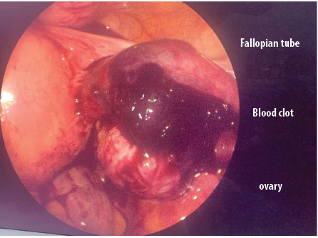 Figure 1: Cornual ectopic pregnancy with blood clot over the ovary. The fallopian tube appears normal.
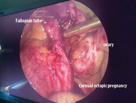 Figure 2: Cornual ectopic pregnancy with blood clot over the ovary and normal looking fallopian tube.
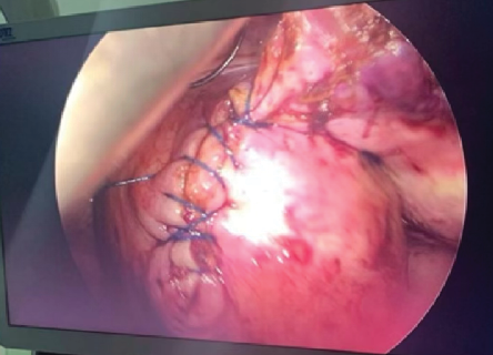 Figure 3: The suture line after closing the resected wedge of the right cornua.
She had a smooth postoperative period and was discharged on the third day post-surgery. Her human chorionic gonadotropin (hCG) level became negative on day 25 post-operation, and the patient did not require any further treatment or medication.
Discussion
Cornual ectopic pregnancy occurs when the embryo embeds itself within the interstitial section of the fallopian tube.2 This rare and dangerous condition requires prompt diagnosis and intervention. Diagnosis can be challenging because the initial presentation can mimic a normal intrauterine pregnancy.3 Hemorrhagic shock has been reported in approximately one-quarter of the patients.4
The causative risks for cornual ectopic pregnancy include assisted reproductive technologies, a history of salpingectomy or other tubal surgical procedures, the presence of a rudimentary horn, prior reproductive tract infection, previous tubal pregnancy, and the development of adhesions within the proximal intratubal area.5 Others include endometriosis, advanced maternal age, and smoking.6
Transvaginal ultrasound is the mainstay of diagnosis.7 Ultrasonographic indications for cornual ectopic pregnancy include a gestational sac that is distinct from the uterine cavity, coupled with an empty uterine cavity. A thin layer of endometrium (< 5 mm) encircling the gestational sac is another indication. Notably, an echogenic line is seen in the central endometrial cavity extending toward the gestational sac. Monitoring the variations in blood hCG levels also helps diagnose cornual ectopic pregnancy, where hCG levels vary more slowly than in intrauterine pregnancy.
Early ultrasonographic diagnosis is essential for successful laparoscopic management, which involves removal of the ectopic pregnancy and hemostasis of the cornua. Two techniques are proposed: (1) cornuotomy and (2) cornual wedge resection, [Figure3] along with ipsilateral salpingectomy. The literature does not indicate any significant difference between the outcomes of the two. The likelihood of blood loss, postoperative complications, and alteration of postoperative fertility seem similar in both. Irrespective of the technique, salpingectomy is technically unavoidable in most cases. Post surgical risk factors include interstitial pregnancy and uterine rupture in subsequent pregnancies.8
The surgical approaches for managing the ectopic pregnancy include cornuostomy (making an incision in the cornua and extracting the pregnancy); cornual evacuation (emptying the contents of the cornual region); cornual area resection (removing of a section of the cornual area); and cornual wedge resection (often conducted alongside an ipsilateral salpingectomy). To minimize blood loss during the procedure, injecting vasopressin around the location of the ectopic pregnancy is recommended.
Careful postoperative monitoring is essential. Subsequent pregnancies should be monitored via early ultrasonography to ascertain the fetal position and the integrity of the surgical site. Women who underwent conservative surgical therapy seem more susceptible to repeat ectopic pregnancies due to the persistence of deeply embedded viable trophoblastic tissue. Therefore, postoperative hCG level should be monitored until it become undetectable.5
Medical management with methotrexate may be considered in selected cases.9 In a prospective observational study in the UK, 17 women with cornual pregnancy were treated with single-dose intramuscular methotrexate. A second dose was given if the hCG levels had not fallen by 15%. All women with an initial hCG value of < 5000 mIU were treated successfully with single-dose methotrexate, but almost all women with an initial hCG > 5000 mIU required two doses.10
The risks of medical treatment include uterine rupture and catastrophic hemorrhage. It may also not be the optimal choice for ectopic pregnancy with fetal heartbeat.11 While several experts have recommended a multiple-dose methotrexate regimen for interstitial/cornual pregnancies, there is no clear evidence for or against this approach.12
Kim et al,13 reported a procedure with 100% success rate involving ultrasound-guided local injection of methotrexate, resulting in immediate cessation of fetal heart activity. The time taken for the resolution of the ectopic pregnancy was similar whether potassium chloride or methotrexate was used. Several studies have also documented the utilization of laparoscopy for locally administering methotrexate into cornual pregnancies.14
An emerging minimally invasive alternative to surgery is selective uterine artery embolization. This involves injecting an embolic agent into the uterine arteries to block the blood supply to the cornual area and the ectopic pregnancy, causing its resolution.15
Conclusion
Cornual ectopic pregnancy is a rare but potentially life-threatening condition that requires prompt diagnosis and management. A high index of suspicion, early ultrasound, and selection of treatment modality are key to favorable outcomes.
Disclosure
The authors declare no conflicts of interest. Written consent was obtained from the patient for publication of this case report.
references
- Walid MS, Heaton RL. Diagnosis and laparoscopic treatment of cornual ectopic pregnancy. Ger Med Sci 2010 Jul;8:Doc16.
- 2. Sargin MA, Tug N, Ayas S, Yassa M. Is interstitial pregnancy clinically different from cornual pregnancy? A case report. J Clin Diagn Res 2015 Apr;9(4):QD05-QD06.
- 3. Dagar M, Srivastava M, Ganguli I, Bhardwaj P, Sharma N, Chawla D. Interstitial and cornual ectopic pregnancy: conservative surgical and medical management. J Obstet Gynaecol India 2018 Dec;68(6):471-476.
- 4. Hoang BT, Whitaker DW. Ruptured left cornual ectopic pregnancy: a case report. Cureus 2023 Jul;15(7):e41449.
- 5. Varun N, Nigam A, Elahi AA, Jain A. Cornual ectopic pregnancy: laparoscopic management step by step. BMJ Case Rep 2018 Mar;2018:bcr2017223998.
- 6. Jacob L, Kalder M, Kostev K. Risk factors for ectopic pregnancy in Germany: a retrospective study of 100,197 patients. Ger Med Sci 2017 Dec;15:Doc19.
- 7. Winder S, Reid S, Condous G. Ultrasound diagnosis of ectopic pregnancy. Australas J Ultrasound Med 2011 May;14(2):29-33.
- 8. Moawad NS, Dayaratna S, Mahajan ST. Mini-cornual excision: a simple stepwise laparoscopic technique for the treatment of cornual pregnancy. JSLS 2009;13(1):87-91.
- 9. Dhar H, Hamdi I, Rathi B. Methotrexate treatment of ectopic pregnancy: experience at nizwa hospital with literature review. Oman Med J 2011 Mar;26(2):94-98.
- 10. Ray A, Gaur A, Kumari S. Predictors of successful medical management with methotrexate in unruptured tubal ectopic pregnancy. Cureus 2022 Nov;14(11):e31923.
- 11. Wang S, Beejadhursing R, Ma X, Li Y. Management of caesarean scar pregnancy with or without methotrexate before curettage: human chorionic gonadotropin trends and patient outcomes. BMC Pregnancy Childbirth 2018 Jul;18(1):289.
- 12. Murray H, Baakdah H, Bardell T, Tulandi T. Diagnosis and treatment of ectopic pregnancy. CMAJ 2005 Oct;173(8):905-912.
- 13. Kim YR, Moon MJ. Ultrasound-guided local injection of methotrexate and systemic intramuscular methotrexate in the treatment of cesarean scar pregnancy. Obstet Gynecol Sci 2018 Jan;61(1):147-153.
- 14. Al-Kharusi L, Gowri V, Al-Sukaiti R, Al-Ghafri W, Rao K. Submyomatous cornual pregnancy: managed surgically after failed medical management. Sultan Qaboos Univ Med J 2011 Aug;11(3):399-402.
- 15. Simon P, Donner C, Delcour C, Kirkpatrick C, Rodesch F. Selective uterine artery embolization in the treatment of cervical pregnancy: two case reports. Eur J Obstet Gynecol Reprod Biol 1991 Jul;40(2):159-161.