There are many structural variations in the organs of the abdominal cavity; however, those connected to the ligament attachment of the liver are crucial from a therapeutic standpoint. One of these is the falciform ligament, which connects the liver to the umbilical ring by extending from the anterior abdominal wall to the diaphragm.1 Previous research has indicated that there are structural differences between adults and children in the falciform ligament's thickness, stretch, and development.2 Adult falciform ligaments have a mean length and thickness of about 9.9 cm and 6 mm, respectively, according to earlier studies. Additionally, the obliterated umbilical vein is enclosed by this ligament.3 Falciform ligament fixation may not occur if the peritoneum fails to merge with the umbilical vein. In the pediatric age group, this anomaly has been linked to newborn intestinal obstruction and internal herniation.
Case Report
We report a case of a 24-year-old male patient who presented with intermittent upper abdominal pain with dyspepsia for the last six months. On ultrasound, he had gallbladder stones with a normal dimension, including the diameter of the common bile duct. Hepatic functions tests results were within the normal range. He was planned for laparoscopic cholecystectomy. During surgery, the pneumoperitoneum could not be created via the Veress needle, and the pressure was high during insufflation. Therefore, Hasson technique was opted for. After the introduction of the telescope, a diagnostic laparoscopy was performed, and everything was normal except the insufficiently formed falciform ligament, which was not dividing the liver into two halves as it normally does. The liver was unsupported on the undersurface of the anterior abdominal wall, and a thick, bulky, fat-laden band of the umbilical vein extended from the umbilicus to the liver, making a congenital band and mainly located at the umbilical region [Figure 1]. The liver extended from the right side of the abdomen to the left without any separation by the falciform ligament [Figure 1]. Consent was obtained from the patient.
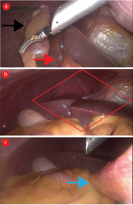 Figure 1: (a) Umbilical vein band extending up to the undersurface of the liver (red arrow) and retracted gall bladder (black arrow). (b) Showing the liver completely unsupported against the anterior abdominal wall due to the absence of attachment of the falciform ligament to the anterior abdominal wall (red rhombus). (c) A thick fat-laden band of umbilical vessels extending from the umbilicus to the liver without extended falciform to the abdominal wall (blue arrow).
Discussion
The double-layered falciform ligament physically divides the liver into right and left segments. This ligament connects the lower edge of the diaphragm to the liver's front-to-back surface. The falciform ligament runs along the anterior abdominal wall from the parietal peritoneum to the visceral peritoneum over the liver surface. It divides the upper part of the supra-mesocolic gaps into two subphrenic spaces.4 Torsion or twisting of the fatty appendageal pockets of the said ligament is extremely rare and known as intra-abdominal focal fat infarction, which clinically resembles other acute surgical conditions.5 The ligament is a crucial reference point for abdominal and gallbladder surgeries. It hides the hepatic vein, which drains the liver into the inferior vena cava.
Usually, the ligament is mainly attached to the anterior abdominal wall, well away from the umbilicus, but in this case, it was not; rather, the thick and bulky end of the ligament was in the umbilicus region, which pos difficulty while creating the pneumoperitoneum. During the difficulty creating of pneumoperitoneum, the palmar point approach or open Hasson technique was the alternative, safer way of creating pneumoperitoneum, which is well described in the literature, and we opted for the latter.
The round ligament, which enters the umbilicus at the free end of the falciform ligament, also acts as an important landmark of the anterior abdominal wall.6 The umbilical vein endures as a solid, band-like structure. Small bowel herniation is the most frequent internal hernia and has been linked to this anomaly in 0.2% of cases.7 In 1960, a baby with pyloric stenosis, an anomalous umbilical vein caused intestinal obstruction, according to a case study by Hoffbert and Strachman.8
Few articles recommend removing the umbilical vein if it is found accidentally because of the risk of intestinal obstruction or volvulus in the future. We did not do this because the umbilical vein helps support the liver, the patient had no episodes of intestinal obstruction, and the patient's preoperative symptoms and ultrasonography of the abdomen pointed only to gallbladder stones.
Conclusion
If there is any difficulty during the creation of pneumoperitoneum for laparoscopy, surgeons should keep this abnormal falciform ligament attachment in mind and opt for another way of creating pneumoperitoneum (i.e., Hasson technique or Palmar’s point entry).
Disclosure
The authors declared no conflicts of interest.
references
- Ozkececı ZT, Ozsoy M, Celep B, Bal A, Polat C. A rare cause of acute abdomen: an isolated falciform ligament necrosis. Case Rep Emerg Med 2014;2014:570751.
- 2. Han SY. Variation in falciform ligament with pheumoperitoneum. J Can Assoc Radiol 1980 Sep;31(3):171-173.
- 3. Lakdawala M, Chaube SR, Kazi Y, Bhasker A, Kanchwala A. Internal hernia through an iatrogenic defect in the falciform ligament: a case report. Hernia 2009 Apr;13(2):217-219.
- 4. Indiran V, Dixit R, Maduraimuthu P. Unusual cause of epigastric pain: intra-abdominal focal fat infarction involving appendage of falciform ligament - case report and review of literature. GE Port J Gastroenterol 2018 Jun;25(4):179-183.
- 5. Justaniah AI, Scholz FJ, Katz DS, Scheirey CD. Perigastric appendagitis: CT and clinical features in eight patients. Clin Radiol 2014 Dec;69(12):e531-e537.
- 6. Garbar V, Newton BW. Anatomy, abdomen and pelvis: falciform ligament. StatPearls [Internet]. Treasure Island (FL): StatPearls Publishing; 2023.
- 7. Wiseman S. Internal herniation through a defect in the falciform ligament: a case report and review of the world literature. Hernia 2000;4(2):117-120.
- 8. Hoffert PW, Strachman J. Intestinal obstruction due to aberrant umbilical vein and hypertrophic pyloric stenosis. Case in a two-week-old infant. Arch Surg 1960 Dec;81(6):890-892.