Cetuximab is a monoclonal antibody that selectively acts on epidermal growth factor receptors (EGFR) by blocking ligand binding and preventing the conformational change of the receptor required for dimerization. EGFRs play a major role in normal cellular processes in epithelial, mesenchymal, and neuronal tissues. Antibodies such as cetuximab may trigger some form of skin toxicity in 80–95% of patients.1 It usually presents as an acne-like papulopustular rash with dry itchy skin, mainly on the face and upper trunk. Less common presentations include seborrheic dermatitis-like eruptions, paronychia, desquamation, pruritus, male pattern alopecia, hypertrichosis, and trichomegaly.1 Although the mechanism is unclear, it is known to be associated with the high EGFR expression that cetuximab promotes.2
Case Report
A 55-year-old woman, with a known type II diabetes mellitus controlled by diet, was diagnosed with stage III sigmoid adenocarcinoma. She underwent laparoscopic high anterior resection in 2018, resulting in the removal of a tumor that had grown through the muscularis propria into the subserosa, and into seven regional lymph nodes. She received 12 cycles of 5-fluorouracil, leucovorin, and oxaliplatin chemotherapy. She tolerated chemotherapy well, but developed grade I peripheral neuropathy. One year later, a positron emission tomography and computed tomography (PET-CT) scan showed an increased uptake in the left para-aortic lymph nodes, which increased in size from 1.9 to 2.4 cm, and the standardized uptake value increased from 7 to 10 with the new left supraclavicular lymph node (1.5 cm; standardized uptake value 6.3). A CT-guided biopsy of the para-aortic lymph node showed metastatic adenocarcinoma consistent with metastasis from the colorectal primary. The tumor molecular profile showed proficient mismatch repair proteins. Immunohistochemically, the tumor was HER-2 negative, and wild type for NRAS, KRAS, and RAF oncogenes.
The patient preferred to avoid multi-agent chemotherapy, but agreed to capecitabine with bevacizumab. Despite an initial response, a PET-CT scan after nine cycles showed progression of disease in left supraclavicular lymph nodes, mediastinal, and para-aortic lymphadenopathy with carcinoembryonic antigen = 10 ng/mL (normal < 5 ng/mL). After counseling, the patient agreed to start on a multi-agent regimen, capecitabine, oxaliplatin with cetuximab.
After six cycles, the patient had abnormally excessive growth of facial hair [Figure 1] and was investigated for hirsutism. Her hormone profile indicated normal levels of thyroid-stimulating hormone of 1.8 UI/mL (reference range = 0.35–5.5 UI/mL), testosterone of 0.396 nmol/L (reference range = 0–2.6 nmol/L), prolactin of 163 mlU/L (reference range = 102–496 mlU/L), cortisol random of 175 nmol/L (reference range = 140–690 nmol/L), dehydroepiandrosterone of 2.63 µmol/dL (0.96–6.95 μmol/dL). Her follicle-stimulating hormone level was 56.32 UI/L (reference range = 30 UI/L) and luteinizing hormone was 33.04 UI/L (reference range = 5–25 IU/L premenopause; reference range = 14.2–52.3 IU/L postmenopause), consistent with her perimenopausal status. Upper abdominal contrast-enhanced CT imaging did not show any adrenal lesions.
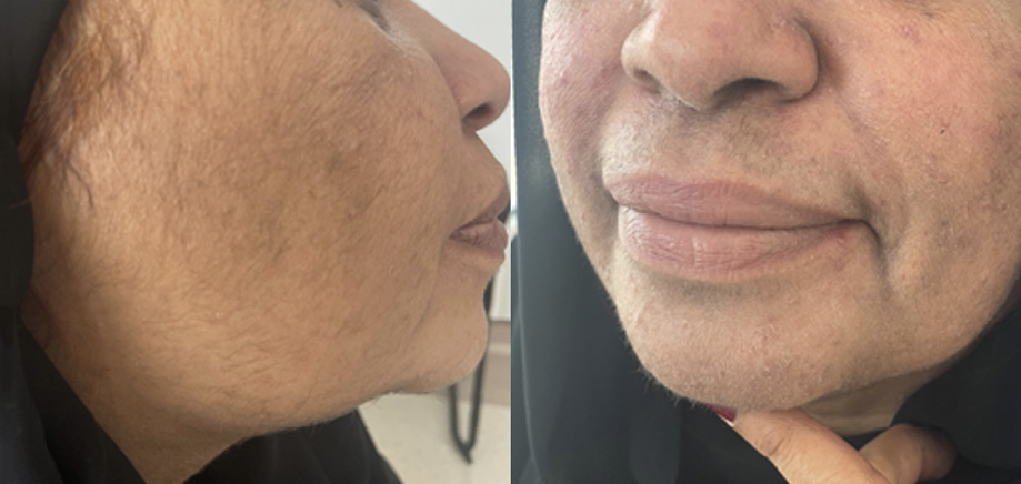 Figure 1: Excessive facial hair growth after treatment with cetuximab.
As the patient was distressed by her cosmetic appearance, cetuximab was withheld for six weeks with close monitoring, which reversed her hirsutism [Figure 2]. She had an excellent response to treatment, as shown by a PET scan after cycle four, and a regimen amounting to almost complete disappearance of the metastatic disease. Written informed consent was obtained from the patient.
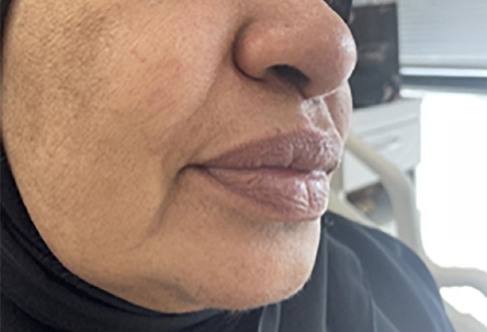 Figure 2: Facial hair regression after pausing cetuximab therapy.
Discussion
Hypertrichosis is a rare, cosmetically distressing side effect of cetuximab. Hair growth typically occurs on the face, scalp, and upper back, and can be permanent or temporary. A woman in Turkey developed hypertrichosis, trichomegaly, and androgenic alopecia following treatment with cetuximab and chemotherapy for RAS-wild-type metastatic colon cancer.3 Montagut et al,4 reported abnormal hair growth and acneiform follicular eruption in a 66-year-old male patient treated with cetuximab and carboplatin in a second-line setting for supraglottic squamous cell carcinoma with resolution of skin toxicity upon cetuximab discontinuation. Vano-Galvan et al,5 reported hair growth in the scalp and eyelashes of a bald hundred-year-old man after cetuximab treatment for squamous cell carcinoma of the scalp. EGFRs are widely distributed in skin tissue, and therefore, the disruption of signaling is involved in the initiation of the anagen phase of the hair cycle, resulting in dysregulation of hair growth.
FIRE-3 (AIO KRK0306), a randomized clinical trial with FOLFIRI and cetuximab, investigated the regimen for potential skin toxicity, acneiform rash, nail changes, desquamation, and dry skin. It showed clinical predictors of good response, but no cases of hypertrichosis were reported among the 199 patients because it is a rare condition.6 On the other hand, an observational study of 116 patients with metastatic colon adenocarcinoma who received anti-EGFR, noted papulopustular rash and xerosis, and considered these as clinical predictors of good response.7
The impact of hypertrichosis on the prognosis of colon cancer is not well understood due to under-reporting. It did not seem to have any impact on our patient who responded very well to the anticancer regimen.
Conclusion
Cetuximab-induced hypertrichosis is rare but can be cosmetically distressing, and is severely underreported in the literature, perhaps because it is seen as a minor cosmetic problem that resolves itself. It is important to give the patient counseling support to overcome any emotional distress, even pausing the treatment till the cosmetic symptoms reverse.
Disclosure
The authors declare no conflicts of interest.
references
- 1. Fakih M, Vincent M. Adverse events associated with anti-EGFR therapies for the treatment of metastatic colorectal cancer. Curr Oncol 2010 Jul;17 Suppl 1(Suppl 1):S18-S30.
- 2. Hu JC, Sadeghi P, Pinter-Brown LC, Yashar S, Chiu MW. Cutaneous side effects of epidermal growth factor receptor inhibitors: clinical presentation, pathogenesis, and management. J Am Acad Dermatol 2007 Feb;56(2):317-326.
- 3. Turker S, Cilbir E, Karacin C, Altinbas M. Hypertrichosis, trichomegaly, and androgenic alopecia related to cetuximab treatment. J Cancer Res Ther 2020;16(3):690-692.
- 4. Montagut C, Grau JJ, Grimalt R, Codony J, Ferrando J, Albanell J. Abnormal hair growth in a patient with head and neck cancer treated with the anti-epidermal growth factor receptor monoclonal antibody cetuximab. J Clin Oncol 2005 Aug;23(22):5273-5275.
- 5. Vano-Galvan S, Ríos-Buceta L, Ma DL, Fernández-Chacón C, Viera JC, Jaén P. Cetuximab-induced hypertrichosis of the scalp and eyelashes. J Am Acad Dermatol 2010 Mar;62(3):531-533.
- 6. Holch JW, Held S, Stintzing S, Fischer von Weikersthal L, Decker T, Kiani A, et al. Relation of cetuximab-induced skin toxicity and early tumor shrinkage in metastatic colorectal cancer patients: results of the randomized phase 3 trial FIRE-3 (AIO KRK0306). Ann Oncol 2020 Jan;31(1):72-78.
- 7. Jaka A, Gutiérrez-Rivera A, López-Pestaña A, del Alcázar E, Zubizarreta J, Vildosola S, et al. Predictors of tumor response to cetuximab and panitumumab in 116 patients and a review of approaches to managing skin toxicity. Actas Dermosifiliogr 2015;106(6):483-492.