A 51-year-old male presented with an eight-year history of gradually progressive skin lesions, initially noted as gray-brown papules on the buttocks that progressively expanded symmetrically to involve the entire gluteal region. The patient denied a history of prior dermatologic conditions affecting the buttocks or elsewhere. Although the lesions were non-pruritic, the patient reported discomfort when sitting due to pressure exerted by the lesions.
Physical examination revealed symmetrical, verrucous, gray-brown plaques localized on the bilateral buttocks, accompanied by satellite lesions [Figure 1a]. A skin biopsy from the affected buttock was performed. The histopathological evaluation demonstrated marked hyperkeratosis and incomplete cornoid lamella, with minimal loss or reduction of the granular cell layer distributed along the length of the biopsy specimen [Figure 1b].
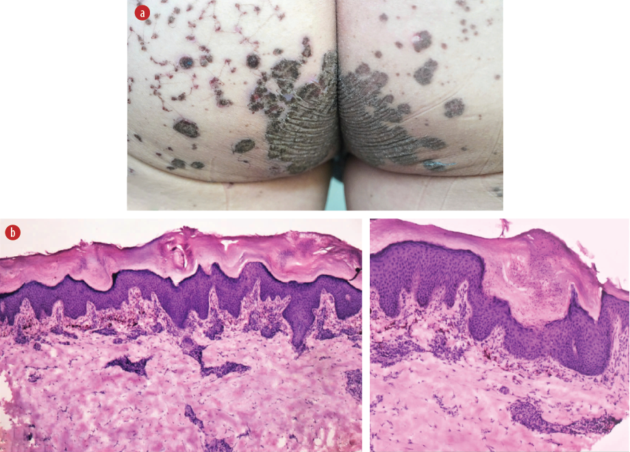 Figure 1: (a) Clinical image showing symmetrical, gray-brown, verrucous, non-pruritic lesions on the buttocks, with surrounding satellite lesions. (b) Histopathological image of a biopsy specimen showing marked hyperkeratosis and incomplete cornoid lamella, with minimal loss or reduction of the granular cell layer distributed along the length of the specimen. The epidermis exhibits irregular hyperplasia with
scattered inflammatory cells and pigment incontinence in the dermis. Hematoxylin and eosin staining, magnification = 20 ×.
Based on clinical presentation and histopatho-logical findings, a diagnosis of porokeratosis ptychotropica (PP) was established. The patient underwent CO2 laser ablation. However, these interventions resulted in minimal clinical improvement. Subsequently, the patient expressed acceptance of his condition in its current state and declined further therapeutic interventions. Written informed consent for the publication of this paper was obtained from the patient.
PP is a rare variant of porokeratosis, characterized by symmetrical, verrucous, erythematous to gray-brown plaques, often accompanied by pruritus. These lesions predominantly affect the gluteal folds, though satellite papules may extend to adjacent regions. PP exhibits a marked male predominance, with onset occurring at any age, but most commonly in middle-aged individuals. The disease progresses gradually, evolving from small papules into large plaques.1
Histopathological examination is diagnostic, revealing cornoid lamella distributed longitudinally along the length of the biopsied lesion, with variable angles and occasional alignment parallel to the sectioned surface. Differential diagnoses may include inverse psoriasis, chronic intertrigo, or lichen simplex chronicus. Due to its rarity, PP is often misdiagnosed clinically without histopathological confirmation.2
Treatment remains highly challenging, compounded by the condition’s rarity. Case reports describe frequent therapeutic resistance to established modalities, though isolated successes have been documented.3
Our patient exhibited clinical features consistent with PP, including male gender, middle age, characteristic verrucous plaques with symmetrical gluteal distribution, and refractoriness to CO2 laser ablation. Notably, unlike typical PP cases, our patient did not report pruritus but experienced discomfort when sitting due to pressure on the lesions. The patient’s refusal of further treatment underscores the ongoing therapeutic challenges in managing PP.
references
- 1. Jin Y, Wu T, Xuan X, Cao J, Chen S, Huang C. Porokeratosis ptychotropica: case reports and literature review. Ann Dermatol Venereol 2025 Mar;152(1):103327.
- 2. Veasey JV, Dalapicola MC, Lellis RF, Campaner AB, Manzione T da S, Rodrigues MC de FS. Porokeratosis ptychotropica: a rare manifestation with typical histological exam. An Bras Dermatol 2016 Jul-Aug;91(4):496-498.
- 3. Ryoo YW, Kim Y, Yun JM, Kim SA. Porokeratosis ptychotropica: a case report. J Yeungnam Med Sci 2023 Oct;40(4):423-425.