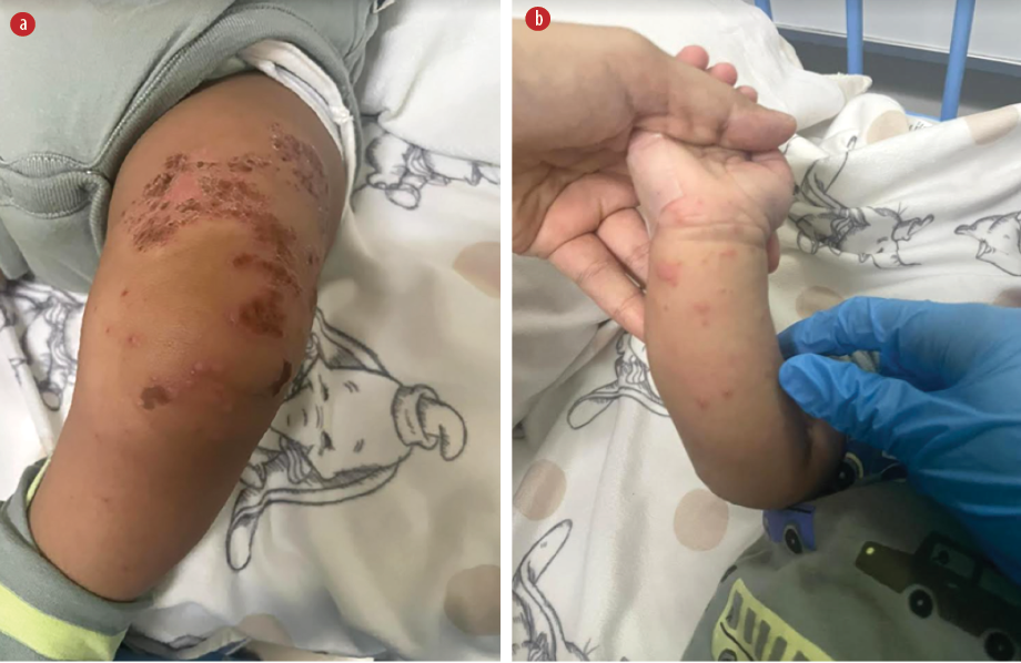
A one-year-old infant, with a known history of glucose-6-phosphate dehydrogenase deficiency and eczema, presented to our emergency room with a one-day history of fever, runny nose, and an exacerbation of eczema along with new skin lesions. The skin exhibited characteristics of pruritic erythematous vesicles with a clear discharge, distributed across the flexor and extensor surfaces of the upper and lower limbs [Figure 1], as well as on the face and neck.
 Figure 1: (a) Dried-out crusted vesicles overlying eczematous plaques with few active intact vesicles over the knee. (b) Active vesiculopapular skin lesions over the arm.
Additional manifestations included tactile fever, diminished oral intake, and reduced activity without evident central nervous system involvement. There were no reported contacts with sick individuals. The patient had previously received two doses of amoxicillin for the skin lesions at a private hospital with no significant improvement. No urticaria or angioedema were observed. Respiratory, gastrointestinal, musculoskeletal, and urinary systems revealed no abnormalities. Laboratory findings indicated microcytic hypochromic anemia with predominant lymphocytosis, a C-reactive protein level of 12 mg/L, and respiratory viral panel positivity for rhinovirus and enterovirus RNA.
Questions
- What is the diagnosis?
- How do you rule out other possible differential diagnoses?
- How would you manage this infant?
Answers
- Eczema coxsackium (EC).
- By detecting herpes simplex virus and varicella-zoster virus by polymerase chain reaction (PCR)from the skin lesions to rule out the possibility of eczema herpaticum (EH) and a bacterial swab culture looking for secondary bacterial infections.
- EC has a self-limiting course, requiring hydration and, if necessary, antipyretics. In addition, optimizing eczema care that involves the use of moisturizers and topical steroids, if needed.
Discussion
Upon admission, the provisional diagnosis of EH was made, and the patient was commenced on fluid hydration along with intravenous acyclovir at a dose of 250 mg/m2 thrice daily. Skin lesion swabs tested positive for enterovirus and negative for varicella-zoster virus and herpes simplex virus using PCR, supporting the diagnosis of EC. Bacterial culture from a skin lesion swab revealed no bacterial growth. Consequently, intravenous acyclovir was discontinued. There was spontaneous resolution of the lesions, which healed and crusted without further deterioration. He was discharged on the third day of admission in a stable condition.
Atopic dermatitis is a prevalent allergic skin disorder among children, posing an increased susceptibility to recurrent bacterial and viral infections such as EH and EC when inadequately controlled.1 The term ‘eczema coxsackium’ was first introduced during a coxsakievirus A6-associated enterovirus outbreak in North America between 2011–2012.2,3 EC is caused by coxsackie viruses, which are part of the genus enterovirus in the Picornaviridae family.1,2,4 It is more common in children, though recent cases in adults have been reported.1 The manifestations of EC in children include oral ulcers and vesiculobullous lesions in the soles and palms similar to hand, foot, and mouth disease. In addition, areas exhibiting active atopic dermatitis like flexures, extensor, and the buttocks may develop vesiculopapular or vesiculobullous lesions similar to our patient.1,2 Differential diagnosis includes bullous impetigo, EH, and varicella-zoster infection.2,4 Despite being a self-limiting disease, EC’s clinical significance lies in its potential misidentification as EH or bullous impetigo, leading to unnecessary administration of acyclovir and antibiotics.1,3,4
In such instances, performing enterovirus PCR on skin lesions helps confirm the diagnosis, thereby preventing/minimizing unnecessary antimicrobial therapy.1,4 In our case, enterovirus PCR on the lesions swab confirmed the diagnosis. Clinical suspicion of EC arose due to the patient’s coryzal symptoms and the detection of enterovirus RNA in his respiratory secretions.
Disclosure
The authors declared no conflicts of interest. Written consent was obtained from the patient’s mother.
references
- 1. Ong PY, Leung DY. Bacterial and viral infections in atopic dermatitis: a comprehensive review. Clin Rev Allergy Immunol 2016 Dec;51(3):329-337.
- 2. Mathes EF, Oza V, Frieden IJ, Cordoro KM, Yagi S, Howard R, et al. “Eczema coxsackium” and unusual cutaneous findings in an enterovirus outbreak. Pediatrics 2013 Jul;132(1):e149-e157.
- 3. Ganguly S, Kuruvila S. Eczema coxsackium. Indian J Dermatol 2016;61(6):682-683.
- 4. Su HJ, Chen CB. Eczema coxsackium. Med J Aust 2021 Nov;215(9):403-403.