Epstein-Barr virus-positive diffuse large B-cell lymphoma, not otherwise specified (EBV+ DLBCL, NOS is a newly recognized aggressive B-cell lymphoma associated with chronic EBV infection.1 The World Health Organisation (WHO) recognizes a pathologic spectrum ranging from a monomorphic pattern with a predominance of large cells to a more common polymorphous subtype characterized by scattered large cells amidst a reactive background.2,3 The large cells may appear as centroblasts, immunoblasts, or as Hodgkin and Reed-Sternberg (HRS)-like cells. Montes-Moreno et al,4 described three morphologic subgroups: a large cell subtype composed of numerous large cells, a second one showing HRS-like large cells resembling classic Hodgkin lymphoma (cHL), and a third subtype resembling T-cell/histiocyte-rich large B-cell lymphoma showing only a few or no HRS-like cells.
Composite lymphoma (CL) is defined as two or more distinct lymphoid neoplasms that occur in the same site.5,6 CL is very rare, representing only 1–4.7% of all lymphoma cases.7 It can occur in various combinations, including composite B-cell lymphoma, composite B- and T-cell lymphoma, and composite HL and non-HL (NHL).6 It can develop synchronously or metachronously.8–10 A diffuse large B-cell lymphoma developing metachronously in nodular lymphocyte-predominant HL is the commonest type of CL.10 In some cases of CL, polymerase chain reaction amplification and sequence analysis of the immunoglobulin heavy chain gene have demonstrated common clonal origins.11,12 The latter is thought to be the common precursor of cells that are genetically identical but morphologically different due to different transformation events.8 Metachronously developed cases may be due to complications from previous chemotherapy associated with immune dysregulation.
We report an unusual case of peripheral T-cell lymphoma (PTCL), NOS that seemed to develop metachronously in the patient with EBV+ DLBCL, NOS with HRS-like cells.
Case Report
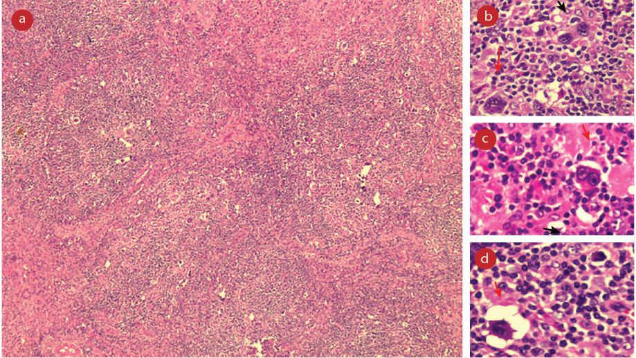
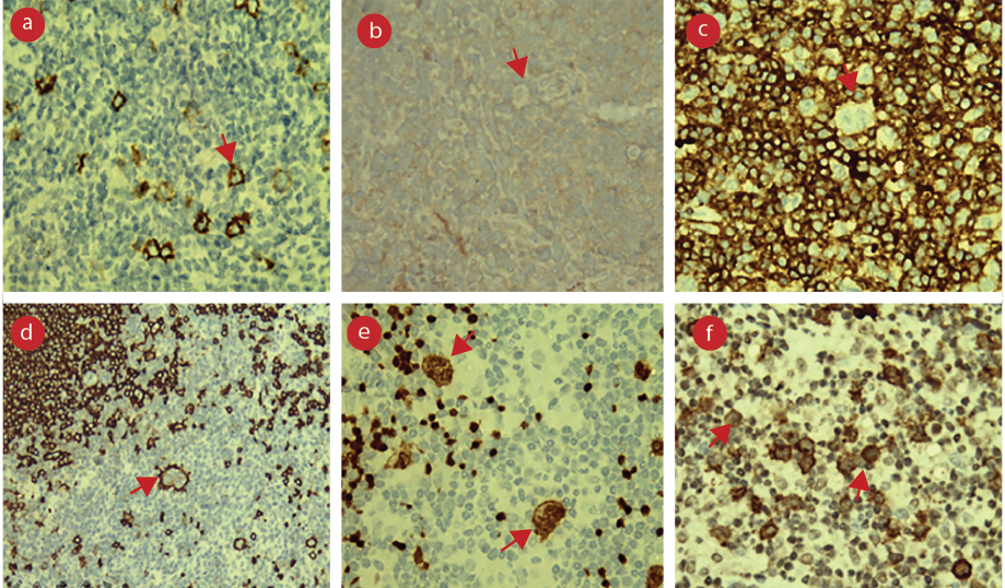
A 70-year-old hypertensive male presented with fatigue and cough with minimal expectoration for three months, not associated with fever or night sweats. He also reported a history of undocumented weight loss. Physical examination revealed enlarged axillary lymph nodes and mild splenomegaly. Hemogram and peripheral blood smear examination showed normochromic anemia with leukopenia and mild thrombocytopenia. Serum lactate dehydrogenase and uric acid levels were mildly elevated. Radiological imaging confirmed splenomegaly and additionally revealed the presence of enlarged para-aortic and peripancreatic lymph nodes. Bone marrow examination, conducted in view of pancytopenia, yielded normal results. Histopathological examination of the left axillary lymph node showed features of reactive lymphoid hyperplasia. Laparoscopic para-aortic lymph node biopsy revealed a lymph node 3 × 2 cm in size with a homogenous, grey-white cut surface. Microscopic sections showed small lymphocytes arranged in a nodular pattern with many scattered large mononuclear, multilobed, and multinucleated cells having abundant cytoplasm and vesicular nuclei with prominent eosinophilic nucleoli. A few lacunar cells, mummified cells, and clusters of histiocytes were also seen [Figure 1]. The large cells were positive for cluster of differentiation (CD) 30, CD20, and paired box 5, but negative for leukocyte common antigen , CD3, and CD15. A mixture of CD20 and CD3-positive lymphocytes formed the background population. A second panel of immunostains performed showed diffuse strong positivity for EBV-latent membrane protein 1, multiple myeloma 1, and B cell-specific octamer-binding protein-1, weak positivity for octamer-binding protein 2, and fascin negativity in the large cells. MIB-1 proliferation index was 80% [Figure 2]. Epstein-Barr encoding region in situ hybridization was not done due to logistic reasons. A final impression of EBV+ DLBCL, NOS was rendered.
 Figure 1: (a) Para-aortic lymph node biopsy showing small lymphocytes arranged in nodular pattern with many scattered large cells (hematoxylin and eosin stain (H&E) = 100 ×), (b) large mononuclear cells (black arrow) and multinucleated giant cells (red arrow) (H&E = 400 ×), (c) classic Reed-Sternberg like cells (red arrow) (H&E = 400 ×), (d) a few lacunar cells (red arrow) and mummified cells (black arrow). The background showing small lymphocytes (H&E = 400 ×).
 CD: cluster of differentiation; LCA: leukocyte common antigen; PAX5: paired Box 5; EBV: epstein-Barr virus; LMP1: latent membrane protein 1.
Figure 2: (a) Para-aortic lymph node biopsy showing CD30 positive large cells (CD30 = × 400), (b) the large cells are seen negative for CD15 (CD15 = 400 ×), (c) the large cells seen negative for LCA (LCA = 400 ×), (d) the large cells are seen positive for CD20 (CD20 = 400 ×), (e) the large cells are positive for PAX5 (PAX5 = 400 ×), (f) the large cells are positive for EBV-LMP1 (EBV-LMP1 = 400 ×).
Positron emission tomography (PET) scan showed F-18 fluorodeoxyglucose (FDG)-avid multiple supra and infradiaphragmatic lymph nodes, spleen, and marrow spaces of axial skeleton (Ann Arbor Stage IV). Quantitative polymerase chain reaction for EBV conducted on blood detected 19 959 copies/mL. Tests ruled out immunodeficiency and autoimmune diseases. The patient was planned for R mini cyclophosphamide, vincristine, doxorubicin, and prednisone 21 × 6 cycles. After three cycles, the patient attained complete FDG PET metabolic response and molecular complete remission. However, as he also developed severe pneumocystis carinii pneumonia and severe renal failure, chemotherapy was discontinued, and rituximab maintenance was initiated.
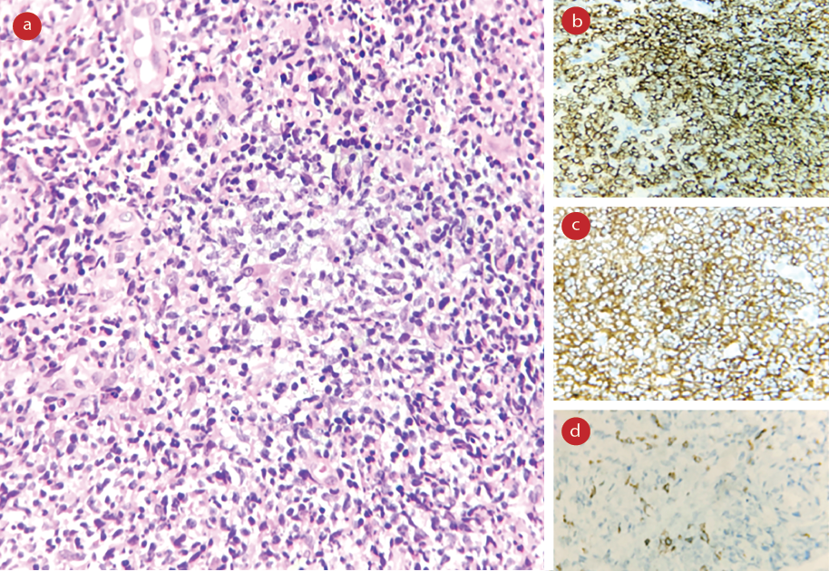
A year and a half later, the patient presented with a three-day history of low backache and abdominal pain. Ultrasonogram revealed borderline splenomegaly and multiple intra-abdominal enlarged lymph nodes with perinodal fat stranding. Whole body PET scan imaging revealed FDG-avid right axillary and deep pectoral, para-aortic, aortocaval, retrocaval, mesenteric, and iliac nodes. Trucut biopsy of the axillary and para-aortic lymph nodes showed a dense infiltrate composed of lymphoid cells, histiocytes, plasma cells, and neutrophils. The lymphoid cells were intermediate-sized with scant to moderate cytoplasm, clumped to vesicular nuclei with irregular to clefted nuclear contours, and inconspicuous nucleoli. A few ill-formed histiocytic collections were also seen [Figure 3]. Special stains for acid-fast bacilli and fungal organisms were non-contributory. Immunohistochemical stains showed that the intermediate-sized lymphoid cells were positive for CD3, CD5, and CD4, and were negative for CD7, CD8, CD10, CD20, CD56, CD30, paired box 5, multiple myeloma 1 , BCL6, and EBV latent membrane protein 1. Ki-67 proliferation index was about 50%.
 Figure 3: (a) Right axillary lymph node biopsy showing dense infiltrate composed of lymphoid cells, histiocytes, plasma cells, and neutrophils. The lymphoid cells were intermediate-sized with scant to moderate cytoplasm, irregular to clefted nuclear contours, and inconspicuous nucleoli.(Haematoxylin and eosin = 400 ×), (b) the atypical cells were positive for cluster of differentiation (CD) 3 (CD3 = × 400), (c) positive for CD4 (CD4 = 400 ×), and (d) negative for CD8 (CD8 = 400 ×).
The histomorphological and immunohistochemical findings were suggestive of PTCL, NOS. T-cell receptor gamma chain gene rearrangement assay confirmed the clonal nature of the T cell population. The patient was started on chemotherapy with cyclophosphamide, vincristine, and prednisolone protocol; however, he succumbed to his illness.
Discussion
EBV+ DLBCL, NOS is a distinct lymphoid neoplasm recognized in the World Health Organization classification of lymphoid neoplasms (4th edition, 2016) and is associated with a poor response to chemotherapy and short survival.1 It was previously designated as EBV+ DLBCL of the elderly as most patients tended to be elderly. Of late, it has been reported over a wide age range and is more common among Asian and Latin American patients compared to Western patients.13,14 Involvement can be nodal or extranodal, the most common extranodal sites being lungs and gastrointestinal tract.14 In some cases, the neoplastic component consists of a variable number of large transformed cells on a background of a mixed population of small lymphocytes, plasma cells, histiocytes, and eosinophils, imparting a morphologic overlap with cHL. When the background reactive component is rich in small lymphocytes and histiocytes, the so-called polymorphic pattern, resembling T-cell/histiocyte-rich large B-cell lymphoma, is the most common pattern in young patients.15 The monomorphic pattern can be distinguished from EBV- DLBCL with ancillary studies only. The neoplastic cells are typically positive for the pan-B-cell antigens with an activated B-cell immunophenotype.14 CD30 is usually positive with occasional CD15 coexpression, but lacks other phenotypic features typical of cHL. The disease has an aggressive course, even when treated with combination chemotherapy.16
CL is an uncommon lymphoid malignancy with a variety of patterns. The most common types are B-cell NHL composite with HL or two composite B-cell NHLs of different types.6 CL consisting of B-cell and T-cell neoplasms are extremely rare, constituting < 1% of all lymphoid malignancies, and the pathologic features of this type have not been well characterized.17 Among the reported cases of this subtype, the T-cell neoplasms were most often PTCL, NOS whereas the B-cell components were diverse with DLBCL being the most frequently followed by marginal zone lymphoma and other low-grade B-cell lymphomas, HL, and plasmacytoma. Most of these cases were of nodal origin, whereas in a minority of cases, they arose in extranodal tissues.
Though the exact pathogenesis of CL remains unclear, a few mechanisms have been hypothesized.18 One possible etiology of CL is the viral-induced transformation of two or more separate neoplastic clones. This oncogenic activity has been observed most frequently in EBV in immunocompromised patients. EBV-related large B-cell lymphoma has been described in association with PTCL and angioimmunoblastic T-cell lymphoma (AITL) in a simultaneous or sequential occurrence.19 It has been thought that EBV antigens expressed in host B-cells may stimulate T-cell proliferation and eventually induce neoplastic transformation of T-cells, via a process of clonal selection. Another hypothesis is immunodeficiency associated with T-cell malignancy predisposes the patient to EBV infection which causes clonal transformation of B-cells in the host.20
This case report adds to the knowledge of the natural history of EBV+ DLBCL, NOS which is a newly described entity. Further studies are required to understand the pathogenesis of metachronous EBV+ DLBCL, NOS and PTCL.
Conclusion
This report presents a noteworthy case of EBV+ DLBCL, NOS, which initially presented as intra-abdominal lymphadenopathy with metachronous development of PTCL, NOS. Being a newly recognized entity, the course of this rare disease needs further study.
Disclosure
The authors declared no conflicts of interest. The institutional ethics committee waived the consent requirement [RAJH/A/2021/004].
references
- 1. Swerdlow SH, Campo E, Pileri SA, Harris NL, Stein H, Siebert R, et al. The 2016 revision of the World Health Organization classification of lymphoid neoplasms. Blood 2016 May;127(20):2375-2390.
- 2. Nakamura S, Jaffe ES, Swerdlow SH. EBV-positive diffuse large B-cell lymphoma, not otherwise specified (NOS). In: Swerdlow SH, Campo E, Harris NL, et al, editors. WHO classification of tumours of haematopoietic and lymphoid tissues. Lyon: IARC; 2017. p. 304-306.
- 3. Ok CY, Papathomas TG, Medeiros LJ, Young KH. EBV-positive diffuse large B-cell lymphoma of the elderly. Blood 2013 Jul;122(3):328-340.
- 4. Montes-Moreno S, Odqvist L, Diaz-Perez JA, Lopez AB, de Villambrosía SG, Mazorra F, et al. EBV-positive diffuse large B-cell lymphoma of the elderly is an aggressive post-germinal center B-cell neoplasm characterized by prominent nuclear factor-kB activation. Mod Pathol 2012 Jul;25(7):968-982.
- 5. Kim H, Hendrickson R, Dorfman RF. Composite lymphoma. Cancer 1977 Sep;40(3):959-976.
- 6. Kim H. Composite lymphoma and related disorders. Am J Clin Pathol 1993 Apr;99(4):445-451.
- 7. Thirumala S, Esposito M, Fuchs A. An unusual variant of composite lymphoma: a short case report and review of the literature. Arch Pathol Lab Med 2000 Sep;124(9):1376-1378.
- 8. Amini RM, Enblad G, Sundström C, Glimelius B. Patients suffering from both Hodgkin’s disease and non-Hodgkin’s lymphoma: a clinico-pathological and immuno-histochemical population-based study of 32 patients. Int J Cancer 1997 May;71(4):510-516.
- 9. Dong C, Hemminki K. Second primary neoplasms among 53 159 haematolymphoproliferative malignancy patients in Sweden, 1958-1996: a search for common mechanisms. Br J Cancer 2001 Sep;85(7):997-1005.
- 10. Brown JR, Weng AP, Freedman AS. Hodgkin disease associated with T-cell non-Hodgkin lymphomas: case reports and review of the literature. Am J Clin Pathol 2004 May;121(5):701-708.
- 11. Bellan C, Lazzi S, Zazzi M, Lalinga AV, Palummo N, Galieni P, et al. Immunoglobulin gene rearrangement analysis in composite hodgkin disease and large B-cell lymphoma: evidence for receptor revision of immunoglobulin heavy chain variable region genes in Hodgkin-Reed-Sternberg cells? Diagn Mol Pathol 2002 Mar;11(1):2-8.
- 12. Bräuninger A, Hansmann ML, Strickler JG, Dummer R, Burg G, Rajewsky K, et al. Identification of common germinal-center B-cell precursors in two patients with both Hodgkin’s disease and non-Hodgkin’s lymphoma. N Engl J Med 1999 Apr;340(16):1239-1247.
- 13. Beltran BE, Castillo JJ, Morales D, de Mendoza FH, Quinones P, Miranda RN, et al. EBV-positive diffuse large B-cell lymphoma of the elderly: a case series from Peru. Am J Hematol 2011 Aug;86(8):663-667.
- 14. Gibson SE, Hsi ED. Epstein-Barr virus-positive B-cell lymphoma of the elderly at a United States tertiary medical center: an uncommon aggressive lymphoma with a nongerminal center B-cell phenotype. Hum Pathol 2009 May;40(5):653-661.
- 15. Nicolae A, Pittaluga S, Abdullah S, Steinberg SM, Pham TA, Davies-Hill T, et al. EBV-positive large B-cell lymphomas in young patients: a nodal lymphoma with evidence for a tolerogenic immune environment. Blood 2015 Aug;126(7):863-872.
- 16. Stuhlmann-Laeisz C, Borchert A, Quintanilla-Martinez L, Hoeller S, Tzankov A, Oschlies I, et al. In Europe expression of EBNA2 is associated with poor survival in EBV-positive diffuse large B-cell lymphoma of the elderly. Leuk Lymphoma 2016;57(1):39-44.
- 17. Suefuji N, Niino D, Arakawa F, Karube K, Kimura Y, Kiyasu J, et al. Clinicopathological analysis of a composite lymphoma containing both T and B-cell lymphomas. Pathol Int 2012 Oct;62(10):690-698.
- 18. Mokhtar NM. Review article composite lymphoma. J Egypt Natl Canc Inst 2007 Sep;19(3):171-175.
- 19. Zettl A, Lee SS, Rüdiger T, Starostik P, Marino M, Kirchner T, et al. Epstein-Barr virus-associated B-cell lymphoproliferative disorders in angloimmunoblastic T-cell lymphoma and peripheral T-cell lymphoma, unspecified. Am J Clin Pathol 2002 Mar;117(3):368-379.
- 20. Xu Y, McKenna RW, Hoang MP, Collins RH, Kroft SH. Composite angioimmunoblastic T-cell lymphoma and diffuse large B-cell lymphoma: a case report and review of the literature. Am J Clin Pathol 2002 Dec;118(6):848-854.