|
Abstract
Kirner`s deformity or dystelephalangy is a rare entity which presents with painless, progressive, bilateral radiovolar curving of the terminal phalanges of the little fingers. It is a clinicoradiological diagnosis. Herein, we present a case where the patient was being treated as having a fracture of the distal phalanx because of misdiagnosis of Kirner`s deformity. Given the rarity of the deformity, we believe it useful to present our case report as a contribution to the literature.
Keywords: Kirner`s deformity; Fifth; Little finger; Radio-volar angulation.
Case Report
A 13-year-old boy was referred to the Radiodiagnosis department for X-ray of bilateral hands from the Orthopedic outpatient department. He presented with bilateral deformities of the little fingers noticed by him three years back. He was being treated by a local practitioner as a case of fractured distal phalanges, but the patient consulted our college because he felt the deformity was gradually progressing. The deformity was not associated with pain, swelling, or redness. There was no history of any local infection in the little fingers. He was otherwise healthy. Also, no history of similar deformity in any one of his siblings or family members was revealed.
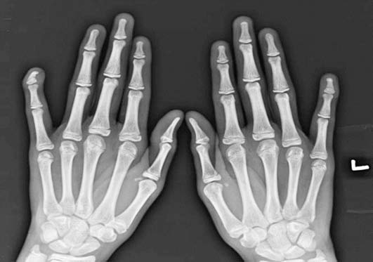
Physical examination of bilateral little fingers showed palmar and radial curving of the distal phalanges, more on the right side. There was no associated tenderness or swelling seen. The nails of the affected fingers were also curved in the volar direction. At the distal interphalangeal (DIP) joint, range of extension was evidently restricted. His routine blood examinations were normal. Rheumatoid factor and C-reactive protein were also negative. X-ray of bilateral hands showed ventral and radial angulation of the terminal phalanx relative to the middle phalanx with an apparent overgrowth of epiphysis on the palmar surface and a tiny bony spur, which projected distally to fit into a groove in the basal part of the shaft. Physeal plate also appeared widened with sharply narrowed and sclerosed diaphysis. (Fig. 1)
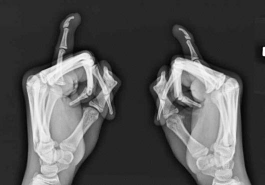
There was no extension into the DIP joint. Lateral view showed palmar bending of the diaphysis of distal phalanx (Fig. 2). Treatment modalities were discussed with the parents. As the deformity was painless, observation with periodic follow up was chosen as the modality of treatment.

Figure 1: X-ray of bilateral hands (AP view) shows ventro-radial angulations of the terminal phalanx with an apparent overgrowth of epiphysis on the palmar surface and a tiny bony spur.

Figure 2: X-ray of bilateral little finger (lateral view) shows palmar bending of the diaphysis of distal phalanx.
Discussion
Kirner`s deformity or dystelephalangy was first described by Kirner in 1927.1 It is a rare but usually bilaterally occurring bony deformity. Literature concerning this deformity is sparse as only a few cases have been reported in the medical journals.
Incidence of the deformity has been reported to be 1/410 by David and Burwood with a higher incidence noted among the Japanese. The deformity is usually sporadic but may be inherited as an autosomal dominant trait with incomplete penetrance with 2:1 female to male ratio. Deformity is bilateral in most cases with right-sided dominance seen in the unilateral cases.2,3
Etiopathogenesis of Kirner’s deformity is still not well understood, with hypotheses being juvenile osteomalacia, aseptic necrosis,4 and osteochondrosis of possible vascular origin.5 More distal insertion of the flexor digitorum profundus tendon along the palmar surface has also been proposed as a cause.6 However, systemic causes seem unlikely.
Clinically, Kirner`s deformity is characterized by shortening of the terminal phalanx of the little finger, which is stubby and deflected in a palmar and radial direction, typically described as "eagle-claw-like" by Sugiura,7 with a small, dysmorphic "watch glass" nail. In majority of the cases, it becomes obvious between 8 and 14 years of age.1,2,5 However, in some cases the deformity may be present since birth. Interestingly, in such cases, as opposed to juvenile onset, the deformity is also seen in other members of the family. The deformity progresses over months to years and ceases with closure of the physis. It is rarely associated with pain, redness or swelling. Functional limitations, if any, are usually minimal and confined to playing musical instruments or typing.8
Deformity needs to be differentiated from other similar deformities such as clinodactyly (radial deviation at the DIP joint) and camptodactyly (flexion deformity at the PIP joint).
Association has been reported with musculoskeletal (genu valgus, pes cavus, myositis ossificans, absence of flexor digitorum superficialis tendon in the little finger) and cardiovascular abnormalities. Literature review reveals association of this deformity with syndromes like Turner`s syndrome, Cornelia de Lange syndrome and Silver syndrome.2,9,10
Radiological findings are consistent, diagnostic and usually straightforward as in our case. There is ventro-radial angulation of the terminal phalanx relative to the middle phalanx with an apparent overgrowth of the epiphysis on the palmar surface and a tiny bony spur. A radiolucent nidus of 1-2 mm may also be seen in the terminal tuft. The articulation of epiphysis with the middle phalanx is usually preserved. As the patient’s age progresses, the diaphysis regains its width and trabecular structure, but usually 10-50 degrees of the deformity persists. No spontaneous resolution of the deformity has been reported.
Treatment modalities recommended are observation, splinting and osteotomy.11 Since the deformity usually ceases after physis closure, reassurance is sufficient. Temporary splinting may be of help in painful cases. Carstam and Eiken advised one or more volar osteotomies leaving an intact dorsal periosteal hinge with K-wire fixation for correction of the deformity.8 Surgery is delayed until physeal closure in order to prevent recurrence of the deformity.8,10
Conclusion
Awareness of this rare entity is a must to avoid misdiagnosis, as in our case. Assurance and observation with periodic follow-up is needed in most cases.
Acknowledgements
No conflict of interest and no financial assistance was taken for this case.
|