| How to cite this article
Prathvi S, Leo TF, Jnaneshwari J. Sneaky Pancreatic Head Mass. Oman Med J 2012 Jan; 27(1):70-71.
How to cite this URL
Prathvi S, Leo TF, Jnaneshwari J. Sneaky Pancreatic Head Mass. Oman Med J 2012 Jan; 27(1):70-71. Available from http://www.omjournal.org/fultext_PDF.aspx?DetailsID=201&type=fultext
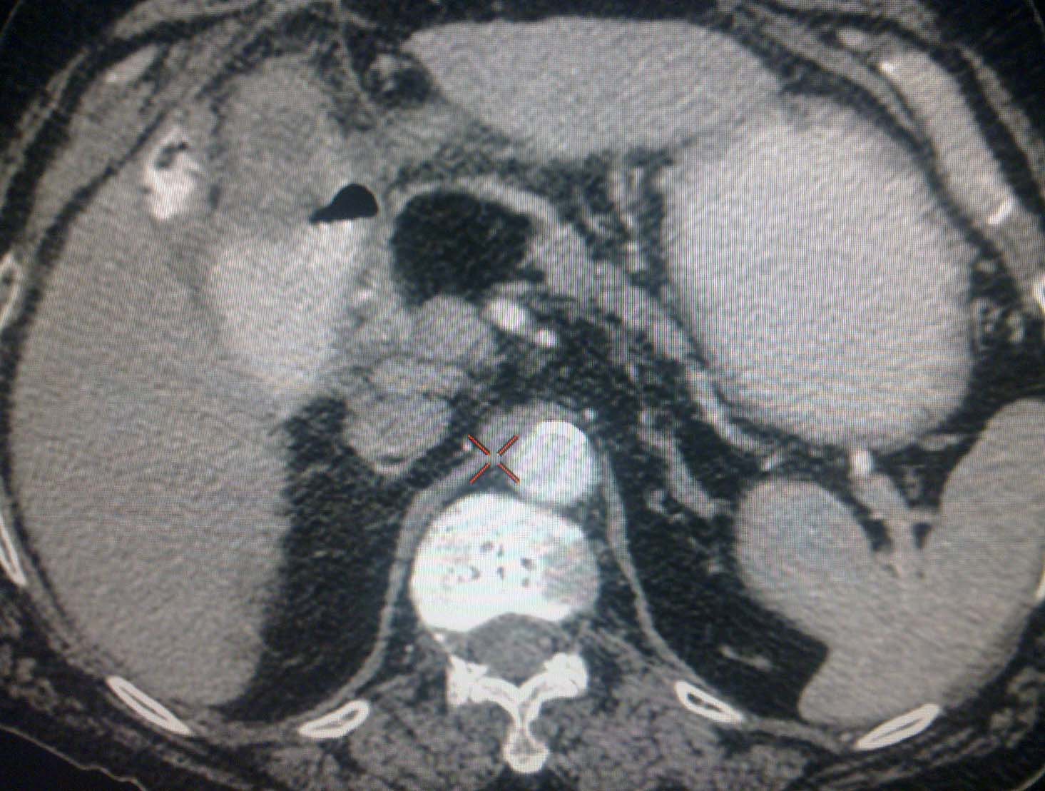
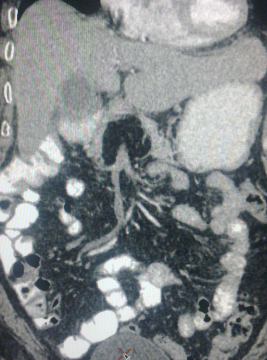
A 60 yrs old male presented to the Father Muller MedicalCollege and Hospital, India, with dyspeptic symptoms. Onphysical examination, epigastric tenderness was present andultrasound revealed an ill defined inhomogeneous hyper echoiclesion in the head of pancreas. Hemogram, serum amylase, serumlipase and liver function tests were all normal. Contrast-enhanced CT showed a homogeneous focal mass measuring about 5 × 6 cmin the pancreatic head, (Figs. 1a and b). The mass was isodensewith fat tissue, with interlobular septa, and without central orperipheral contrast. Upper GI endoscopy revealed mild antralgastritis. The patient improved with antacids.

Figure 1a: Contrast-enhance Computer Tomography.

Figure 1b: Contrast-enhance CT Computer Tomography.
Question
1. What is the diagnosis and management?
Answer
Lipoma of the pancreas
Discussion
Typical CT findings are hypodensity (from -30 to -120 HU)and homogeneity, with no significant contrast enhancement and without infiltration of peri-pancreatic fat. Lipomas appear ashyperechoic on ultrasound with posterior acoustic attenuation, with some instances of hypoechogenicity. MRI is extremely helpful in detecting the presence or absence of macroscopic fat. On T1-weighted images, mature adipose tissue demonstrates high signal intensity and signal drop on fat suppressed sequence; while a T2-weighted image shows variable signal intensity with no enhancement on contrast images.1,2
Pancreatic lipoma is a rare condition usually found incidentally. They are mostly asymptomatic and appear as a well circumscribed, encapsulated homogenous adipose mass within the pancreatic parenchyma. Imaging features of pancreatic lipoma are diagnostic and do not need histopathological evidence.3 In some instances, EUS-FNA is required to differentiate between the benign lipoma and neoplastic lesions, especially lipomatous malignancies.4
There are not many differential diagnosis for pancreatic lipoma on CT scan; few include focal fatty infiltration of the pancreas, teratoma (mature dermoid cyst), and liposarcoma. Histopathological evidence is only needed if there is rapid progressin size to rule out liposarcoma.5 Surgical intervention is done if there are signs of ductal or vessel obstruction and hemorrhage. The condition may be treated with a Whipple procedure, distal pancreatectomy, or by enucleation if the tumors are amenable; if not, palliative by-pass surgery is performed.6,7
Acknowledgements
The authors reported no conflict of interest and no funding was received for this work.
References
1. Karaosmanoglu, Devrim MD, Karcaaltincaba, Musturay MD, Akata, DenizMD, et al. Pancreatic lipoma computed tomography diagnosis of 17 patientsand follow-up. Pancreas. 2008 May; 36:434-6.
2. Katz DS, Nardi PM, Hines J, Barckhausen R, Math KR, Fruauff AA, et al.Lipomas of the pancreas. AJR Am J Roentgenol 1998 Jun;170(6):1485-1487.
3. Temizoz O, Genchellac H, Unlu E, Kantarci F, Umit H, Demir MK.Incidental pancreatic lipomas: computed tomography imaging findings withemphasis on diagnostic challenges. Can Assoc Radiol J 2010 Jun;61(3):156-161.
4. Suzuki R, Irisawa A, Hikichi T, Shibukawa G, Takagi T, Wakatsuki T,et al. Pancreatic lipoma diagnosed using EUS-FNA. A case report. JOP2009;10(2):200-203.
5. Raut CP, Fernandez-del Castillo C. Giant lipoma of the pancreas: case reportand review of lipomatous lesions of the pancreas. Pancreas 2003 Jan;26(1):97-99.
6. Zhan HX, Zhang TP, Liu BN, Liao Q, Zhao YP. A systematic reviewof pancreatic lipoma: how come there are so few cases? Pancreas 2010Mar;39(2):257-260.
7. Salman Monte Z, Ruiz-Cabello Jiménez M. P. Pardo Morenoand P. MontoroMartínez. Lipoma of the pancreas: diagnosis and management of these raretumors. Rev Esp Enferm Dig 2006 Nov;98:11.
|