|
Abstract
Diaphragmatic injury following blunt thoracoabdominal trauma is rare and is usually associated with key radiological features like dependent viscera sign, collar sign, diaphragmatic thickening and defects. It may also be associated with secondary signs like intrathoracic herniation of abdominal viscera. Diaphragmatic crura, which are attached to the upper lumbar vertebra represent prominently thickened folds along the posterior diaphragm, are usually inconspicuous on routine Computed Tomography (CT) scans. We present a case of a young patient who sustained a motor vehicle accident and developed difficulty in breathing. CT scan of the patient revealed bilateral crural hematomas, with splenic and renal lacerations and no other sign of diaphragmatic injury. The patient was operated and blunt diaphragmatic rupture was confirmed at surgery.
Keywords: Diaphragm; Thorax; CT scan.
Introduction
Blunt diaphragmatic ruptures are difficult to diagnose at initial presentation because of non-specific clinical features. Moreover, the radiological findings are usually unsupportive. However, these injuries do not resolve spontaneously; therefore, it warrants timely management. Consequently, a high degree of suspicion should be kept in all cases with thoracoabdominal injuries. They are usually seen in association with other injuries and solitary diaphragmatic injury has never been reported. Few radiological signs have been mentioned in literature but crural hematomas mimicking enlarged lymph nodes have never been described. This case report highlights the importance of this unique radiological sign in early diagnosis of diaphragmatic rupture.
Case Report
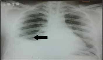
A 24-year-old man was brought to the emergency department after sustaining blunt thoracoabdominal trauma after a motor vehicle injury. On examination, vitals of the patient were stable with mild tachycardia. General examination of the patient was unremarkable and respiratory examination of the patient revealed reduced right sided air entry and fine crepitations were heard along the right lung base. Preliminary routine investigations were normal and the patient's chest x-ray (postero-anterior view in erect position) revealed blunt right costophrenic angle with elevated right hemidiaphragm. The x-ray showed no evidence of rib fractures or pneumothorax. Being hemodynamically stable, the patient was put on conservative management. However, 7 hours after admission, the patient developed severe chest pain that was not relieved by NSAID’s. He did not develop any tachypnea and his oxygen saturation remained normal.
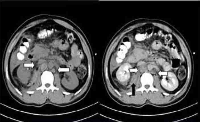
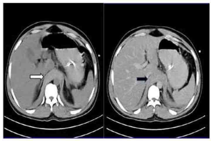
A CT scan of thorax and abdomen was done to elucidate the cause of chest pain. It revealed bilateral hemothoraces and crural hematomas, where the enlarged crura were extending inferiorly in close relation to the abdominal aorta bilaterally simulating retroperitoneal lymphadenopathy. On non-contrast CT (NCCT) images, these lesions reflected diffuse hyperdensity. Additionally, renal and splenic lacerations were encountered with perinephric hematoma and moderate hemoperitoneum.

Figure 1: Chest Radiograph Posteroanterior view of the patient, showing blunt right costophrenic angle with elevated right hemidiaphragm.
The patient was taken for laparotomy, where blunt tear of right hemidiaphragm was confirmed with no evidence of adhesions or herniation of the bowel loops. No bowel injury was noted and the spleen and the kidney were left untouched. The defect was sutured and the patient had an uneventful post-operative course, and subsequently the patient was discharged. Follow up X-ray of the patient two days after surgery revealed no detectable abnormality.

Figure 2: Non contrast and post contrast CT scan of the patient showing well defined nodular lesions in para-aortic location, with hyperdensity on non-contrast images representing crural hematoma (White arrows), also evident is right perinephric hematoma (Black arrows).

Figure 3: Axial non-contrast and post contrast CT scan of the patient shows thickened diaphragmatic crura, which appear hyperdense on NCCT images (White arrow) representing crural hematoma.
Discussion
Diaphragm is a musculofibrous septum that separates thorax from abdominal cavity. The overall incidence of diaphragmatic injuries in patients sustaining blunt trauma ranges between 1-8%,1 out of which 90% of cases are secondary to motor vehicle accidents.2,3 There is an overall left sided predilection, which is affected almost 3 times more than right side.3,4 The possible proposed explanation for left sided preponderance is provided by the buffering effect of liver on right side and an area of potential weakness along the posterolateral aspect on left side.
The overall mortality due to blunt diaphragmatic injuries is low, but it is a marker of fatal injury as mortality in such patients due to other injuries ranges between 12-42%. The clinical diagnosis of these injuries is extremely difficult and often missed; as the presenting symptoms are non-specific and includes shortness of breath or vague chest pain. In a study conducted by Atoinin F. et al. right post traumatic diaphragmatic injuries are usually missed at initial presentation, with a mean period of 47 days and 15 years.5
Chest radiographs are the initial tool in the diagnosis and have a diagnostic accuracy ranging from 27-73%.6,7 Their sensitivity is enhanced if serial x-rays are done in the first 24 hours of injury.6,7 CT scan remains the pivotal investigation in diagnosis of these injuries. As many as 19 different radiological signs of diaphragmatic injuries have been reported in literature. The most common of these include visualization of diaphragmatic defects, collar sign, dependent viscera sign, diaphragmatic thickening and peridiaphragmatic contrast extravasation. "Collar sign" refers to collar like constriction produced by the torn diaphragm around herniated abdominal viscera. "Dependent viscera sign" occurs due to failure of the normal anchoring effect of intact diaphragm, which prevents direct contact of the abdominal viscera with posterior chest wall in supine position. With its rupture, these viscera fall under effect of gravity along the "dependent" part of chest wall. The overall sensitivity of helical CT in the detection of these injuries varies from 71-100% with a specificity of 75-100%.8,9 However, diaphragmatic rupture in association with crural hematomas has rarely been encountered. Few reports of diaphragmatic thickening have been cited in such patients, where the thickening has been attributed to infolding or curling of the free surface of the diaphragm or partial tear.4 In such cases, other signs of diaphragmatic injury coexisted. The measurement of diaphragmatic thickness should be taken 10 mm off the midline to avoid misinterpretation due to normal variations in the size of diaphragmatic crus.4 This sign is highly sensitive for diaphragmatic injuries with sensitivities reaching almost 100%, but the utility of this sign is limited because it cannot differentiate full thickness ruptures from partial tears. This distinction is important since partial tears can be managed conservatively.4 In addition, hemorrhage can track from injury to other adjacent structures, thus, limiting its accuracy.4
The optimal treatment of these injuries is early surgical repair,9 as they never heal on their own. Further, the thoracoabdominal pressure gradient during respiration may enlarge the defect and can lead to complications such as bowel strangulation and formation of adhesions.
Isolated crural hematomas extending inferiorly in the para-aortic location bilaterally, simulating retroperitoneal lymphadenopathy has never been described in patients with diaphragmatic ruptures. Key features in distinguishing them from retroperitoneal lymphadenopathy were hyperdensity on non-contrast images and no perceptible enhancement on contrast administration. Retroperitoneal nodes enlarge as a result of malignant or inflammatory processes. . On CT, they appear as discrete, spherical structures with variable contrast enhancement. The purpose of this case report is to highlight the importance of crural hematomas in diagnosing diaphragmatic injuries, as delay in diagnosis can lead to life threatening complications.
Conclusion
Diaphragmatic injuries though uncommonly encountered in clinical practice, can be fatal if not treated promptly. Imaging studies are pivotal in making an accurate and timely diagnosis. Numerous radiological signs of diaphragmatic rupture have been mentioned in literature, but crural hematomas have seldom been described. Crural hematomas are non-enhancing spherical structures and may be confused with retroperitoneal lymphadenopathy. A comprehensive knowledge of imaging features coupled with keen observation is required for differentiating between the two entities and making a prompt diagnosis.
Acknowledgements
The authors reported no conflict of interest and no funding was received on this work.
References
1. Chughtai T, Ali S, Sharkey P, Lins M, Rizoli S. Update on managing diaphragmatic rupture in blunt trauma: a review of 208 consecutive cases. Can J Surg 2009 Jun;52(3):177-181.
2. Nchimi A, Szapiro D, Dondelinger RF. Injuries of the diaphragm. In:Dondelinger RF, ed. Imaging and intervention in abdominal trauma. Berlin, Germany: Springer-Verlag, 2004; 205–236
3. Shanmuganathan K, Killeen K, Mirvis SE, White CS. Imaging of diaphragmatic injuries. J Thorac Imaging 2000 Apr;15(2):104-111.
4. Nchimi A, Szapiro D, Ghaye B, Willems V, Khamis J, Haquet L, et al. Helical CT of blunt diaphragmatic rupture. AJR Am J Roentgenol 2005 Jan;184(1):24-30.
5. Atoini F, Traibi A, Elkaoui H, Elouieriachi F, Elhammoumi M, Sair K, et al. Missed right post-traumatic diaphragmatic injuries: a review of six cases. Rev Pneumol Clin 2012 Jun;68(3):185-193.
6. Rodriguez-Morales G, Rodriguez A, Shatney CH. Acute rupture of the diaphragm in blunt trauma: analysis of 60 patients. J Trauma 1986 May;26(5):438-444.
7. Gelman R, Mirvis SE, Gens D. Diaphragmatic rupture due to blunt trauma: sensitivity of plain chest radiographs. AJR Am J Roentgenol 1991 Jan;156(1):51-57.
8. Killeen KL, Mirvis SE, Shanmuganathan K. Helical CT of diaphragmatic rupture caused by blunt trauma. AJR Am J Roentgenol 1999 Dec;173(6):1611-1616.
9. Larici AR, Gotway MB, Litt HI, Reddy GP, Webb WR, Gotway CA, et al. Helical CT with sagittal and coronal reconstructions: accuracy for detection of diaphragmatic injury. AJR Am J Roentgenol 2002 Aug;179(2):451-457.
|