|
Abstract
A 25-year-old male presented with complaints of abdominal pain for the past two months. He was prescribed Buscopan and omeprazole by a general surgeon. It was only when patient started complaining of backache that he was referred to an orthopedic surgeon. On examination of spine, there was tenderness at the upper dorsal spine. A diagnosis of Pott’s disease affecting the upper dorsal spine was made. Magnetic resonance imaging (MRI) confirmed the vertebral changes and showed subligamentous spread of paravertebral masses from D2 to D7. This case illustrates the unusual form in which spine tuberculosis can present. High index of suspicion is necessary for early diagnosis and prompt management. General surgeons should be aware of this atypical presentation of Pott’s disease.
Keywords: Tuberculosis; Spine; Pott’s spine; Abdominal pain.
Introduction
Tuberculosis is the most common of infections worldwide.1 Vertebral tuberculosis is the most common form of skeletal tuberculosis and it constitutes about 50% of all cases of tuberculosis of bones and joints.2 It was first described by Sir Percivall Pott in 1779.3 The most common symptom is backache, and rarely caries spine may be responsible for pain referred to the abdomen resembling appendicitis, cholecystitis, pancreatitis, or renal disease. The diagnosis may be missed with atypical manifestations if the findings are misinterpreted. The importance of recognizing this condition stems from the fact that it has the potential to cause severe neural damage and deformity.
This case presents a patient with Pott’s disease of upper dorsal spine complaining of pain over the lower abdomen. Diagnosis was delayed until the patient started complaining of backache. Conservative management with anti-tubercular chemotherapy yielded good clinical results.
Case Report
A 25-year-old Indian man of low socioeconomic status presented to the outpatient department with lower abdominal pain and weight loss for the past 2 months. He used to cry during episodes of abdominal pain, hold his lower abdomen and inguinal region, and wanted to stay in one place and not move a muscle. It sometimes awakened the patient from sleep. The pain was aggravated with walking/jogging and relieved partially with rest. There was no radiation or migration of pain. There were no bowel/bladder complaints, vomiting, or worm infestation. Initially, the patient presented to a general surgeon where hemogram, serum electrolytes, kidney function test, liver function test, Widal test, serum amylase, and ultrasonography of the abdomen were done. All the investigations were within normal limits and the patient was prescribed Buscopan and omeprazole.
The patient did not get relief from his symptoms and started complaining of upper backache after 3 weeks. Due to the backache, he was not able to do his job and had to frequently take leave from work. He then presented to the outpatient department as above. There was a negative family history for tuberculosis and other pulmonary disease. Physical examination revealed a well nourished, alert, afebrile man. Examination of the head, neck, heart, lungs, abdomen, genitalia, and the lymphatic system revealed no abnormalities. On palpation, tenderness was present in the upper dorsal spine and both paraspinal regions. Tone was increased in lower limbs and power was normal in all four limbs. Knee reflex and ankle reflex were exaggerated. He had extensor plantar response bilaterally and well-sustained ankle and patellar clonus. His blood tests on admission showed a total leukocyte count (TLC) of 4500, raised sedimentation rate (60 mm after 1 hour by Wintrobe’s method), hemoglobin (Hb) of 11.8 g/dL, and normal international normalized ratio (INR), prothrombin time and activated partial thromboplastin time. The radiographs of the dorsal spine were unremarkable. Magnetic resonance imaging (MRI) of dorsal spine showed perivertebral collection from D2 to D7 vertebrae (Fig. 1). Rim-enhancing bilateral paraspinal collections, compatible with abscesses, were seen with craniocaudal and intraspinal extensions. Chest radiograph showed right hilar lymphadenopathy with paracardiac infiltrates suggestive of tuberculosis. ELISA test for HIV I and II antibodies was negative. A diagnosis of Pott’s spine D2-D6 with referred pain to the abdomen was made.
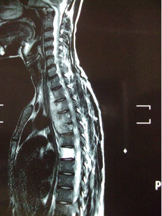
Figure 1: Magnetic resonance imaging (MRI) of dorsal spine showed perivertebral collection from D2 to D6 vertebrae.
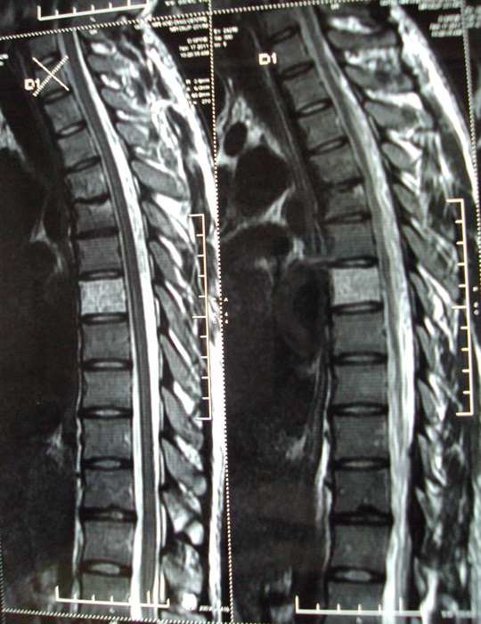
Figure 2: Repeat MRI of dorsal spine showed complete resolution of paraspinal abscess.
The patient was started on anti-tubercular treatment (isoniazid 5 mg/kg; rifampicin 10 mg/kg; pyrazinamide 25 mg/kg; ethambutol 15 mg/kg; along with pyridoxine 10 mg). During his three-week hospital stay, there was a gradual increase in his TLC from 4500 to-9000/cu mm (µl), with a differential shift to marked neutrophilia. Erythrocyte sedimentation rate (ESR) was normal at 19 mm/hr and C-reactive protein (CRP) was mildly prominent at 6.6 mg/L. The standard four-drug therapy of isoniazid (5 mg/kg), rifampicin (10 mg/kg), ethambutol (15 mg/kg) and pyrazinamide (25 mg/kg) was administered at the intensive phase and continued for a total of three months, followed by a three–drug antituberculous treatment (rifampicin/isoniazid/ethambutol) for nine months (maintenance phase). The total duration of antituberculous treatment was 12 months.
At 12-months follow up, the patient was asymptomatic. Repeat MRI of dorsal spine showed complete resolution of paraspinal abscess (Fig. 2). The patient was informed that data concerning the case would be submitted for publication, and written informed consent authorizing radiological examination and photographic documentation was taken.
Discussion
The symptoms of tuberculous bone and joint infections are nonspecific, and the clinical course is often indolent, usually leading to significant delays in diagnosis and resultant bone or joint destruction. Only about 50% of patients with bone and joint tuberculosis have chest radiographs suggestive of tuberculous infection, further obscuring the diagnosis. Tuberculosis of the spine is a rare disease especially in developed countries. Infections of the spine can pose a wide range of problems for both the patient and the surgeon. Tuberculosis of spine has the potential for serious morbidity, including permanent neurologic deficits and severe deformity. Although any part of the spine can be involved, there is a predilection for the lower thoracic and upper lumbar vertebrae.2 In spinal tuberculosis, onset of symptoms is usually insidious and disease progression is slow. The usual presentation includes low grade fever, irritability, weight loss, back pain, kyphosis, abnormal positioning of gait and paraplegia.2,3 Atypical presentations of spinal tuberculosis mimicking malignancy and fracture have been described.4-6
Pande et al. highlighted one of the atypical presentations of tuberculosis of the spine as a herniated lumbar intervertebral disc and a cause of a failed back syndrome.7 These atypical presentations of spinal tuberculosis present a challenge for an appropriate diagnosis and early treatment due to the atypical clinical and radiographic features. The case patient had abdominal pain as an unusual presenting symptom of spinal tuberculosis. Such unusual symptoms may prompt an initial extensive workup for other disease processes. This can lead to serious consequences of a late or missed diagnosis particularly one that is accompanied by irreversible neurological sequelae including paraplegia. Spinal tuberculosis presenting as acute abdomen has been described in some reports, but in these reports, the abdominal pain occurred due to psoas abscess with the etiology being tuberculosis.8 Abdominal ultrasound may pick up hypoechoic lesions suggestive of psoas abscess in 60% of patients but may not be able to identify an underlying etiology.9 MRI have 90% sensitivity and 80% specificity for diagnosing psoas abscess.10 In this case report, the ultrasound of the abdomen was normal and there was no evidence of any abscess even in MRI.
Patients with thoracic spine disease are at particular risk for paraparesis or paraplegia.11 The patient in this case was fortunate that the condition was recognized and managed appropriately. Spinal tuberculosis causing referred pain to the abdomen was not considered in this patient, leading to a delay in the diagnosis. Three theories have been put forward to explain the phenomenon of spinal referred pain: the axon reflex theory, the convergence theory and the hyperexcitability theory. The axon reflex theory suggests that certain primary sensory neurons have bifurcating axons innervating both somatic and visceral targets, leading to confusion as to the source of afferent activity.12 The convergence theory suggests that the afferent nerve fibers from one region converge in the spinal cord with afferent nerve fibres from another region onto a common second order neuron, thereby allowing misinterpretation of the source of pain by the central nervous system.12,13 The hyperexcitability theory suggests that the referred pain occurs through cross connections between second-order neurons supplying the different regions, but only when the input reaches a certain threshold.13,14
Studies done by Kellgren and Feinstein show common patterns of pain referral following irritation of thoracic and lumbar spinal somatic structures.15,16 Usually such radiation occurs following affectation of the lumbar spine, but the presented case is rare in terms of the referred pain which may occur even with the lower dorsal spine affected. Pain is referred outward and downward from its source, in predictable patterns, as far anteriorly as the anterolateral chest and abdomen. The pain is usually felt as deep and dull, or aching, and is diffuse in its distribution. The patient had the typical somatic referred pain due to tuberculous involvement of the D7 vertebra. The anterior abdominal wall is innervated by the intercostal nerves (T7-T12), so the thoracic spine may be a source of abdominal symptoms.
Both visceral sympathetic and somatic nociceptive afferents converge in the same dorsal horn. Also visceral and somatic noxious stimuli may be conveyed in the same spinothalmic tract. Thus pain from the spinal muscles or vertebral bodies may be interpreted as visceral in origin. Jorgensen and Fossgreen compared 39 patients with upper abdominal pain without a demonstrable organic intra-abdominal cause and 28 healthy controls.17 They found that 72% of their patients also had back pain, compared with 17% of controls. The examinations were blinded with regard to the symptoms, 75% of the patients with back pain had vertebral abnormalities shown at the physical examination, pointing to some organic mechanism affecting the spine. Most of the findings were localized to the lower thoracic and thoracolumbar segments, the same ones that innervate the upper gastrointestinal tract.
Spine tuberculosis is a medical disease and anti-tuberculosis drugs have a main role in the recovery and response of patients. The efficacy of anti-tuberculosis drugs and bed rest have been shown in several studies for the treatment of spinal TB in the absence of neurologic deficit and deformity.18,19 Adequate early pharmacological treatment can prevent severe complications and may obviate the need of surgery.20 Combination of rifampicin, isoniazid, ethambutol and pyrazinamide for 3 months followed by a combination of rifampicin, isoniazid and ethambutol for 9 months produced good response in the case of the presented patient. Although tuberculosis comes in the list of differential diagnosis for any clinical presentation, it is unusual to have pain in the abdomen due to spinal tuberculosis. When patients present with persistent or recurrent abdominal pain, the tendency is to consider a visceral source and overlook spine as source of origin. Since spine tuberculosis was not suspected as a cause of abdominal pain, the general surgeon failed to examine the spine nor sought an early consultation from orthopedic surgeon. This led to missing the diagnosis in this case.
These patients are often subjected to variety of procedures/investigations in an attempt to find a cause: simple investigations may give way to more complex and invasive ones in order to find the diagnosis. Failure to find a visceral cause for the pain may lead the general surgeon to label the pain as functional. Clinical picture can sometimes be misleading and should be kept in mind while examining patients with chronic abdominal pain. The general surgeon should keep spinal tuberculosis as differential diagnosis in patients presenting with chronic abdominal pain when no other etiology of pain is found. Missing such a condition can have disastrous outcome for the patients such as paraparesis/paraplegia. Awareness by the general surgeon that abdominal pain may occur as a result of spine tuberculosis can forestall a fruitless search for intra-abdominal pathology. A careful clinical history and physical examination of the spine and being alert to this rare possibility may permit an early diagnosis to be made and appropriate treatment given. Constitutional symptoms such as fever, night sweats, anorexia and weight loss frequently occur in patients with spinal tuberculosis. Surgeons should specifically look for spinal tenderness in such patients and refer them to orthopedic surgeon in case of any suspicion.
Conclusion
Through this case we wish to highlight this unusual presentation of spinal tuberculosis. Also, spinal tuberculosis should be considered in the differential diagnosis and a complete physical and neurological examination should be done in patients presenting with chronic abdominal pain. General surgeons should be aware of this atypical presentation of Pott’s disease. With biological control of the disease by the employment of modern antitubercular drugs, the present day physician can give a better quality of life to the patient,11 provided it is diagnosed early.
Acknowledgements
The authors reported no conflict of interest and no funding was received for this work.
References
1. Al-Rikabi AC, Arafah MA. Tuberculosis of the tongue clinically masquerading as a neoplasm: a case report and literature review. Oman Med J 2011 Jul;26(4):267-268.
2. Tuli SM. Tuberculosis of the spine. In Tuberculosis of the skeletal system 3rd edition. Noida, Jaypee Brothers Medical Publishers, 2004.p. 191-198.
3. Pott P. The chirurgical works of Percivall Pott, F.R.S., surgeon to St. Bartholomew’s Hospital, a new edition, with his last corrections. 1808. Clin Orthop Relat Res 2002 May;(398):4-10.
4. Ringshausen FC, Tannapfel A, Nicolas V, Weber A, Duchna HW, Schultze-Werninghaus G, et al. A fatal case of spinal tuberculosis mistaken for metastatic lung cancer: recalling ancient Pott’s disease. Ann Clin Microbiol Antimicrob 2009;8:32.
5. Chakraborty PP. Deviated tongue: the presenting manifestation of spinal tuberculosis. Indian J Pediatr 2009 Sep;76(9):967-969.
6. Dass B, Puet TA, Watanakunakorn C. Tuberculosis of the spine (Pott’s disease) presenting as ‘compression fractures’. Spinal Cord 2002 Nov;40(11):604-608.
7. Pande KC, Pande SK, Babhulkar SS. An atypical presentation of tuberculosis of the spine. Spinal Cord 1996 Dec;34(12):716-719.
8. Goni V, Thapa BR, Vyas S, Gopinathan NR, Rajan Manoharan S, Krishnan V. Bilateral psoas abscess: atypical presentation of spinal tuberculosis. Arch Iran Med 2012 Apr;15(4):253-256.
9. Lee YT, Lee CM, Su SC, Liu CP, Wang TE. Psoas abscess: a 10 year review. J Microbiol Immunol Infect 1999 Mar;32(1):40-46.
10. Tomich EB, Della-Giustina D. Bilateral psoas abscess in the emergency department. West J Emerg Med 2009 Nov;10(4):288-291.
11. Tuli SM. Results of treatment of spinal tuberculosis by "middle-path" regime. J Bone Joint Surg Br 1975 Feb;57(1):13-23.
12. McMahon SB. Mechanism of cutaneous, deep and visceral pain. In: Wall PD, Melzack R (eds). Textbook of Pain, 3rd ed. Edinburgh (UK), Churchill Livingstone, 1994, p.129-151.
13. Harding G, Yelland M. Back, chest and abdominal pain - is it spinal referred pain? Aust Fam Physician 2007 Jun;36(6):422-423, 425, 427-429.
14. Arendt-Nielsen L, Svensson P. Referred muscle pain: basic and clinical findings. Clin J Pain 2001 Mar;17(1):11-19.
15. Kellgren JH. On the distribution of pain arising from deep somatic structures with charts of segmental pain areas. Clin Sci 1939;4:35-46.
16. Feinstein B, Langton JN, Jameson RM, Schiller F. Experiments on pain referred from deep somatic tissues. J Bone Joint Surg Am 1954 Oct;36-A(5):981-997.
17. Jørgensen LS, Fossgreen J. Back pain and spinal pathology in patients with functional upper abdominal pain. Scand J Gastroenterol 1990 Dec;25(12):1235-1241.
18. Alothman A, Memish ZA, Awada A, Al-Mahmood S, Al-Sadoon S, Rahman MM, et al. Tuberculous spondylitis: analysis of 69 cases from Saudi Arabia. Spine (Phila Pa 1976) 2001 Dec;26(24):E565-E570.
19. Chadha M, Agarwal A, Singh AP. Craniovertebral tuberculosis: a retrospective review of 13 cases managed conservatively. Spine (Phila Pa 1976) 2007 Jul;32(15):1629-1634.
20. Nene A, Bhojraj S. Results of nonsurgical treatment of thoracic spinal tuberculosis in adults. Spine J 2005 Jan-Feb;5(1):79-84.
|