A 42-year-old multiparous woman presented with chronic pelvic pain, predominantly on the right side, with a history of 2–3 episodes of heavy menstrual bleeding. As she had relief after taking tranexamic acid, her management was conservative, but pelvic pain was affecting the quality of her life. X-ray showed a 5 × 5 cm mass in her right pelvis with calcifications. A second woman, 58 years old and postmenopausal, presented with complaints of poor flow of urine, burning micturition, and heaviness in the lower abdomen. X-ray of the kidney, ureter, and bladder (KUB) and ultrasonography of the pelvis were advised. The findings of KUB X-ray were similar to those of the first case.
Question
- What are the most likely diagnoses of cases one and two?
a. Uterine calcified fibroid.
b. Appendicolith.
c. Large vesical calculus.
d. Gossypiboma.
e. Hydatid cyst.
f. Phlebolith.
Answer
a. Uterine calcified fibroid [Figure 1].
c. Large vesical calculus [Figure 2].
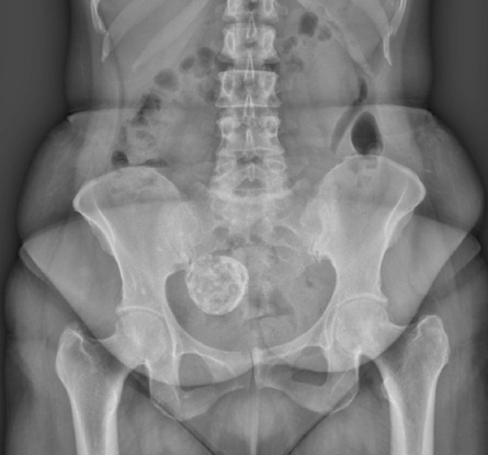 Figure 1: A right-sided pelvic calcified lesion with popcorn appearance. The right uterine wall shows a calcified fibroid.
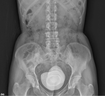 Figure 2: A X-ray image showing a midline stone (large vesical calculus with concentric lamellation) in the pelvis.
The first patient was referred to our department because a pelvis X-ray showed a right-sided calcified mass, a transvaginal scan done for her was suggestive of a 5 × 6 cm calcified fibroid as it had the typical appearance of the echogenic rim with posterior shadowing on the right-sided uterine wall. The patient underwent a total abdominal hysterectomy. The histopathology report indicated calcified leiomyoma.
The second patient had similar findings on KUB X-ray with concentric lamellation in the centre of the pelvis. On ultrasonography, it was found to be an 8.6 × 6.3 cm vesical stone. The patient was managed with suprapubic cystolithotomy. The large stone was taken out [Figure 3] and the bladder was sutured in three layers. The postoperative period was uneventful. The stone was sent for biochemical analysis, which reported a combination of calcium oxalate and calcium phosphate.
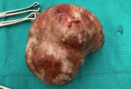 Figure 3: A large vesical calculus after suprapubic cystolithotomy.
Discussion
Uterine fibroids are common in the reproductive age group. They can be asymptomatic or can have variable clinical presentations ranging from heavy menstrual bleeding, dysmenorrhea, to mass per abdomen according to size, shape, and location. Degeneration in fibroids is also common but calcified degeneration is rare with a prevalence of 3–10% of cases.1 The usual ultrasonography findings of uterine fibroids tend to indicate hypoechoic heterogeneous nodules with a smooth regular outline with a predominant occurrence in women in their fourth and fifth decades of life.2 The classical appearance of calcified fibroid on radiography is a popcorn appearance or peripheral rim of calcification with diffuse central calcified areas.3 The presence of calcification in the uterus is a reliable sign of uterine leiomyoma.1
Calcified fibroids are mainly seen in the menopausal age as an aging factor. In a long-standing fibroid, calcified degeneration takes place due to a decrease in blood supply to the uterus. For symptomatic patients with gross fibroid calcification, the treatment of choice is total abdominal hysterectomy with bilateral salpingo-oophorectomy similar to case one. Asymptomatic patients detected incidentally can be managed conservatively. In the literature, cases of calcified fibroids are mainly reported in the postmenopausal age group and can be rarely seen in the reproductive or perimenopausal age group as in case one.
Vesical stone composes almost 5% of the urinary tract calculi.4 Women are at risk for vesical stones due to voiding dysfunction and urinary stasis that can occur secondary to cystoceles, enteroceles, or findings of previous urethral surgery, the rare exception can be any foreign body such as the intrauterine contraceptive device in the bladder which may trigger calcification and eventual stone formation. Small vesical stones are asymptomatic and can be managed easily with transurethral cystolitholapaxy or percutaneous suprapubic cystolitholapaxy, but the large vesical stones require open suprapubic cystotomy requiring a long hospital stay and prolonged catheterization as in case two. It is expected that advances in surgical technology and the ability to downsize without sacrificing effectiveness could eventually render open surgery for stones obsolete.
Though radiographs are not the first investigation of choice for both the above types of pathologies, it is still useful in detecting them and investigating further to reach a diagnosis. Similar radiographic findings can be noted in appendicolith (usually seen in children), which is a piece of firm feces mixed with mineral deposits and can cause appendicitis and may present as a calcified mass on the right side of the pelvis.5
Phleboliths are differentiated from urolithiasis by the presence of a radiolucent center and a ‘comet tail’ sign. Gossypiboma is caused by an introduced foreign body such as a surgical swab retained over a prolonged period, giving a calcified reticulate rind sign in computed tomography.6
Disclosure
The authors declared no conflicts of interest. A written consent was given by the patient's mother.
references
- Tantipalakorn C, Khunamornpong S, Sirilert S, Tongsong T. Popcorn appearance of severely calcified uterine leiomyoma: image-pathological correlation. Diagnostics (Basel) 2023 Jan;13(1):154.
- 2. Kandel P. Calcified fibroid of uterus. [cited 2023 July 13] Available from: https://radiopaedia.org/cases/calcified-fibroid-of-uterus.
- Edzie EK, Dzefi-Tettey K, Brakohiapa EK, Abdulai AB, Kekessie KK, Aidoo E, et al. Assessment of the clinical presentations and ultrasonographic features of uterine fibroids in adult Africans: a retrospective study. Oman Med J 2023 Jan;38(1):e459.
- Schwartz BF, Stoller ML. The vesical calculus. Urol Clin North Am 2000 May;27(2):333-346.
- Kaya B, Eris C. Different clinical presentation of appendicolithiasis. The report of three cases and review of the literature. Clin Med Insights Pathol 2011 Mar;4:1-4.
- Lu YY, Cheung YC, Ko SF, Ng SH. Calcified reticulate rind sign: a characteristic feature of gossypiboma on computed tomography. World J Gastroenterol 2005 Aug;11(31):4927-4929.