|
Abstract
Objectives: To evaluate the significance of P53 and Ki-67 expression as immunohistochemical markers in early detection of premalignant changes in different types of colorectal adenomas. Also, to correlate immunohistochemical expression of the two markers with different clinicopathological parameters including; age, and sex of the patient, type, site, size and grade of dysplasia of colorectal adenomas.
Methods: Forty-seven polypectomy specimens of colorectal adenomas were retrieved from the archival materials of the Gastrointestinal and Hepatic Diseases Teaching Hospital in Baghdad from 2009 - 2010. Four µm section specimens were stained by immunohistochemical technique with Ki-67 and P53 tumor markers. P-values <0.05 were considered statistically significant.
Results: Immunohistochemical expressions of Ki-67 and P53 had a significant correlation with the size and grade of dysplasia in colorectal adenomas. However, there was no significant correlation among the immunohistochemical expression of Ki-67 and P53 with the age and gender of the patient, and the type and site of colorectal adenomas. There was no significant correlation between Ki-67 and P53 expressions in colorectal adenomas. Villous adenomas of colorectum showed a significant correlation with the grade of dysplasia, while there was no significant correlation between size and site of colorectal adenoma with the grade of dysplasia.
Conclusion: High grade dysplasia with significant positive immunohistochemical markers of Ki-67 and P53 could be valuable parameters for selecting from the total colorectal adenoma population, those most deserving of close surveillance in follow-up cancer prevention programs. It is closely linked with increasing age particularly in patients with a large size adenoma of villous component in their histology.
Keywords: Colorectal adenomas; Dysplasia; p53; ki-67 expression; Immunohistochemical expression.
Introduction
Colorectal adenomas are intraepithelial neoplasms that range from small, often pedunculated polyps to large sessile lesions. There is no gender preference, and they are present in nearly 50% of adults by the age of 50. Although adenomatous polyps are not the only precancerous lesions in the colorectum, they are the most common, and they are (especially neoplastic villous type) the main precursors of most of the cancers in this organ.1 While adenomas are less common in Asia, their frequency has risen in these populations as Western diets and lifestyles become more common. Colorectal adenomas are characterized by the presence of epithelial dysplasia. Consistent with their role as precursor lesions of carcinoma, the prevalence of colorectal adenomas correlates with that of colorectal carcinoma and the distribution of adenomas and adenocarcinoma within the colon are similar.
Depending on their characteristics (multiplicity, size, histological features, and grade of dysplasia), these lesions can be associated with a substantial risk of recurrence and the development of advanced neoplastic disease. It has been estimated that 15% of all adenomas measuring >1 cm will progress to carcinomas within 10 years of their detection.2
Ki-67 is a nuclear and nucleolar protein, which is present in cycling cell.3 In contrast to many other cell cycle associated proteins like PCNA (Proliferating Cell Nuclear Antigen), the Ki-67 antigen is consistently absent in quiescent cells and is not detectable during DNA repair processes. Thus, the presence of Ki-67 antigen is strictly associated with cell cycle and confined to the nucleus, suggesting an important role of this structure in the maintenance and /or regulation of the cell cycle.4
The fundamental position of P53 in the cell cycle as the guardian of the genome reflects its central role in the DNA damage response.5 The P53 gene is mutated and deleted in approximately one half of colorectal tumors, leading to inactivation of the P53 protein, and most of the mutations map almost within its DNA binding domains.6 The transcription factor P53 lies at the center of a protein network that controls cell cycle progression (growth arrest in cell cycle stages G1 and G2) and commitment to programmed cell death.7
Several studies have been published evaluating the clinical use of P53 and Ki-67 immunohistochemical expression as predictors of malignant transformation in colorectal adenomas. A significant correlation between the grade of dysplasia and p53 immunoreactivity was observed.8 Ki-67 expression was not related to dysplasia and no correlation was found between P53 and Ki-67 immunoreactivity.8 This study was conducted to evaluate the significance of P53 and Ki-67 expression as an immunohistochemical markers in early detection of premalignant changes in different types of colorectal adenomas. The study also aims to assess the relationship between immunohistochemical expression of these two markers with different clinicopathological parameters such as; age, and sex of patient, and site, type, size, and grade of dysplasia of colorectal adenomas.
Methods
In this retrospective study, forty seven formalin fixed paraffin embedded polypectomy specimens from patients having single colorectal adenoma without concurrent or previous colorectal adenocarcinoma were retrieved from the archival materials of the Gastrointestinal and Hepatic Diseases Teaching Hospital in Baghdad from January 2009 - March 2010. Clinicopathological parameters including the age and gender of the patients, site, size, histological type of the polyp, and grade of dysplasia were obtained from the available histopathological reports.
For each case, one representative section was stained with Hematoxylin and Eosin and the histopathological diagnosis was revised, while two sections were put on positively charged slides and stained immunohistochemically for Ki-67 and P53. The immunohistochemistry select immunophosphatase secondary detection system uses biotin avidin alkaline phosphatase complexed antibodies to detect antimouse IgG in the primary antibody. The alkaline phosphatase activity on the chromogenic substrate results in the deposits of the brown insoluble precipitates at those antigenic sites containing the specific epitopes recognized by the primary antibody. A diffuse brown nuclear stain shows a positive expression for both Ki-67 and P53.9 This positivity was analysed as follows; Ki-67 being a nuclear antigen which is expressed at all stages of the cell cycle, apart from G0, and the anti-Ki-67 antibody, MIB-1, is useful for the analysis of cell proliferation in paraffin wax embedded sections. Ki-67 expression was evaluated semi quantitatively. It was scored by counting at least 1000 cells in 10 high power fields. Every stained nucleus was considered positive, irrespective of intensity. The percentage of positive stained cells was recorded as Ki-67 labeling index (Ki-67-LI).10
P53 is a nuclear antigen. Expression was evaluated semi quantitatively. It was scored by counting at least 1000 cells in 10 high power fields. Every stained nucleus was considered positive, irrespective of intensity. The percentage of positive stained cells was recorded as P53 labeling index (P53-LI).11
For quality control, a negative control had been processed identical to that of the patients sample without addition of the primary antibody for (Ki-67 and P53) to the specimen. For positive control, sections from poorly differentiated colorectal adenocarcinoma patients were stained for Ki-67 and P53.12
Statistical analysis was performed with SPSS 10.01 (statistical package for social sciences), and Excel 2003 programs. Data analysis was done using t- test and chi-square test for tables with frequencies, percentages, mean and standard deviation. Values were considered statistically significant when p<0.05.
Results
A total of 47 tissue samples as paraffin blocks of colorectal polypectomy specimens were included in this retrospective study. Histological examination of Haematoxilin and Eosin stain (H&E) sections confirmed that 28 cases were tubulovillous, 14 cases were villous, and 5 cases were tubular adenomas. The mean age of the patients was 56.78 ± 1.64 (SEM) years, with a range of (29-76) years. Most of patients included in this study were (38) cases in the age group (50-69) years, followed by the age group 40-49 years (5 cases). (Fig. 1)
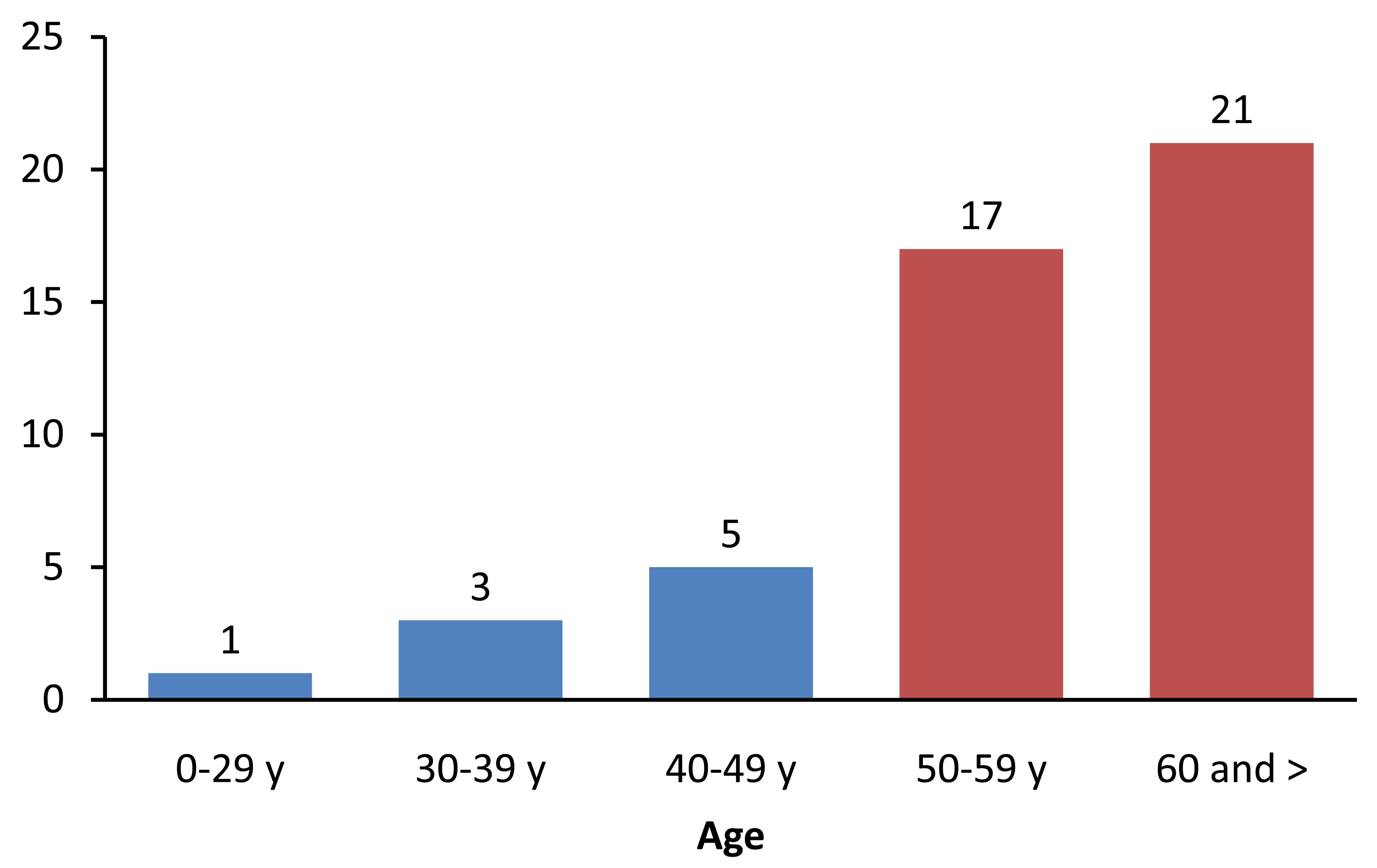
Figure 1: Distribution of Colorectal Adenomas according to Age.
Gender distribution of colorectal adenoma cases, showed male predominance 28 (60%) compared with female 19 (40%), and a male to female ratio of about 1.5: 1.
Regarding the site of colorectal adenomas, the distal site of the colon was the predominant with 25 cases (53.2%), 14 cases (34%) in the proximal site and 8 cases (17%) in the rectum.
The mean size of colorectal adenomas was 1.27 cm ± 0.11; ranging from 0.3 - 3.2 cm. The mean size for Villous adenomas was 1.14 cm; while Tubular adenomas size ranged from 0.8 - 3 cm, with a mean of 1.38 cm. For Tubulovillous adenomas, the size ranged from 0.3 - 3.2 cm, with a mean of 1.4 cm. Relatively, the mean size of different types of adenomas was rather similar; but there is no significant correlation between the size and type of adenomas (p=0.618). (Table 1)
Table 1: Correlation between the Type and the Size of Adenomas.
|
Type of Adenomas
|
No. of
cases
|
Mean
size (cm)
|
SD
|
Min.
size (cm)
|
Max.
size (cm)
|
P
Value
|
|
Villous
|
14
|
1.14
|
0.64
|
0.5
|
2.5
|
0.618
|
|
Tubular
|
5
|
1.38
|
0.95
|
0.8
|
3.0
|
|
Tubulovillous
|
28
|
1.40
|
0.85
|
0.3
|
3.2
|
|
Total
|
47
|
1.32
|
0.79
|
0.3
|
3.2
|
Regarding the grade of dysplasia, there were 27 cases (57%) with low grade dysplasia, and 20 cases (43%) with high grade dysplasia. (Fig. 2)
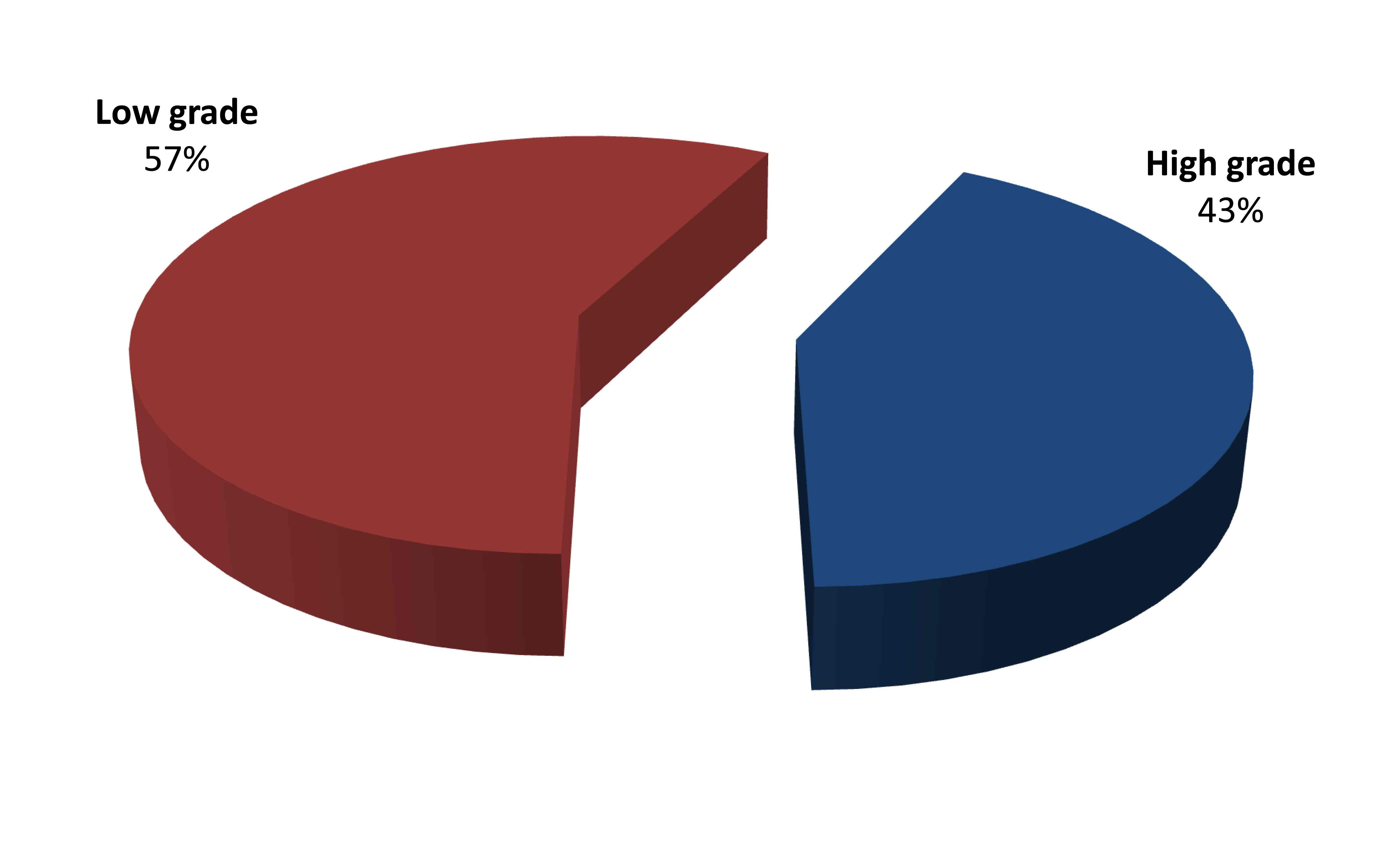
Figure 2: Grade of Dysplasia regardless the type of Colorectal Adenomas.
In Table 2; although p=0.029, there was no significant correlation between the grade of dysplasia and different types of colorectal adenomas as there were 3 cells in this table with a value less than 5.
Table 2: Correlation between Type of Adenomas and Grade of Dysplasia.
|
p= 0.029
|
Types of colorectal adenoma
|
|
Dysplasia
|
Villous
|
Tubular
|
Tubulovillous
|
Total
|
|
High
|
10
|
1
|
9
|
20
|
|
Low
|
4
|
4
|
19
|
27
|
|
Total
|
14
|
5
|
28
|
47
|
There was no significant correlation between the site of colorectal adenomas and grade of dysplasia in colorectal adenomas (p=0.419). (Table 3)
Table 3: Correlation between Site and Grade of Dysplasia in Colorectal Adenomas.
|
p= 0.419
|
Dysplasia
|
|
Site
|
High Grade
|
Low Grade
|
Total
|
|
Distal
|
9
|
16
|
25
|
|
Proximal
|
8
|
6
|
14
|
|
Rectum
|
3
|
5
|
8
|
|
Total
|
20
|
27
|
47
|
Immunohistochemical study There was a highly significant correlation between the immunohistochemical expression of P53 and the size of colorectal adenomas (p=0.001, correlation coefficients (R) = 0.438), and the immunohistochemical expression of Ki-67 and the size of colorectal adenomas regardless the type of the adenoma (p=0.002, R=0.422). (Fig. 3)
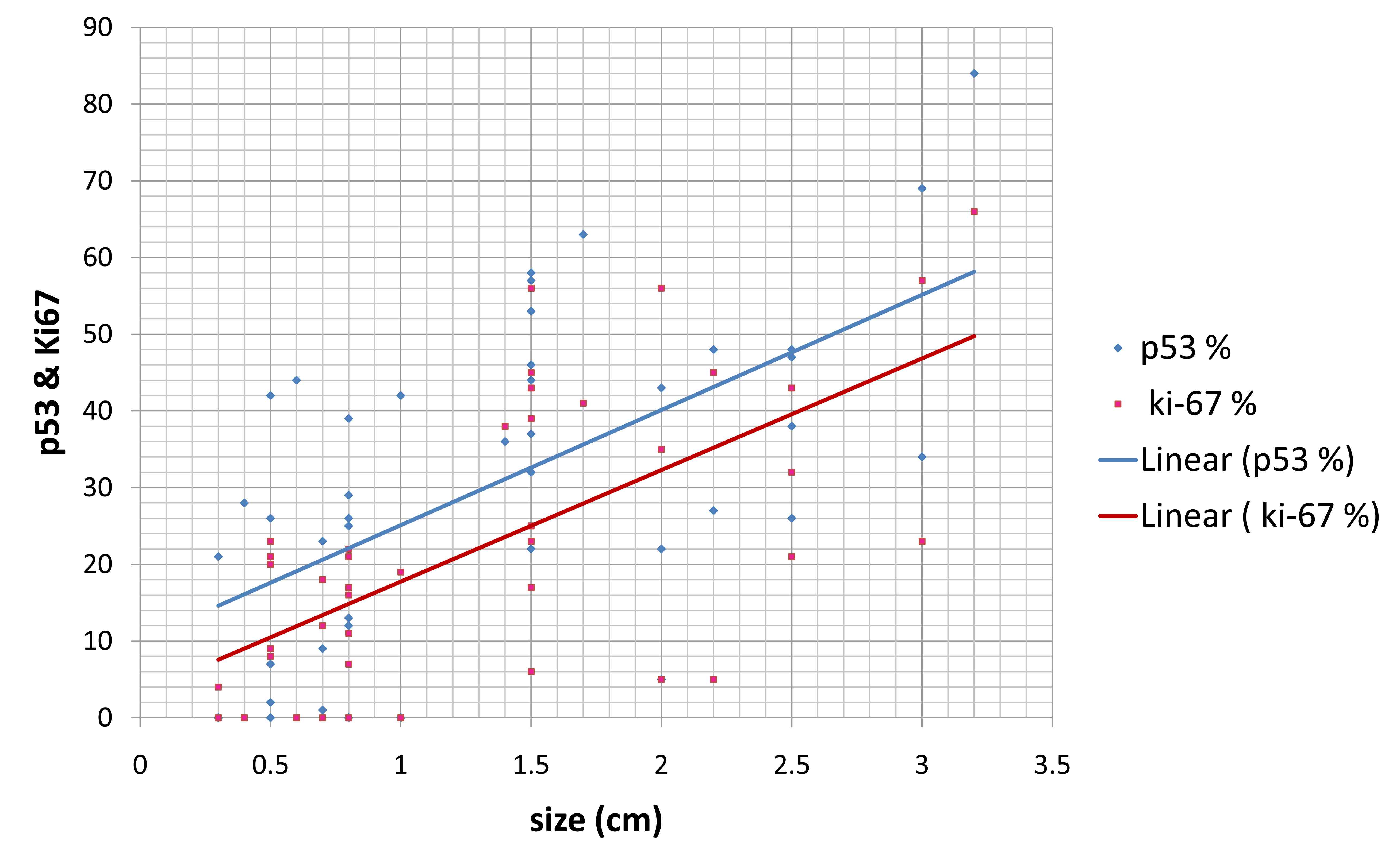
Figure 3: Correlation of P53 and ki-67 expressions with Size of Adenomas.
Also, the immunohistochemical expression of both markers conveyed a highly significant correlation with the grade of dysplasia. (Table 4, Figs 4, 5, 6, 7)
Table 4: Expressions of both Markers according to Grade of Dysplasia.
|
Immunohisto- chemical Markers
|
Grade of Dysplasia
|
Number
of cases
|
Mean of expression
|
SEM
|
p value
|
|
Ki-67
|
high
|
20
|
33.20
|
20.475
|
0.006
|
|
low
|
27
|
13.93
|
9.782
|
|
P53
|
high
|
20
|
44.45
|
18.355
|
0.002
|
|
low
|
27
|
19.19
|
15.059
|
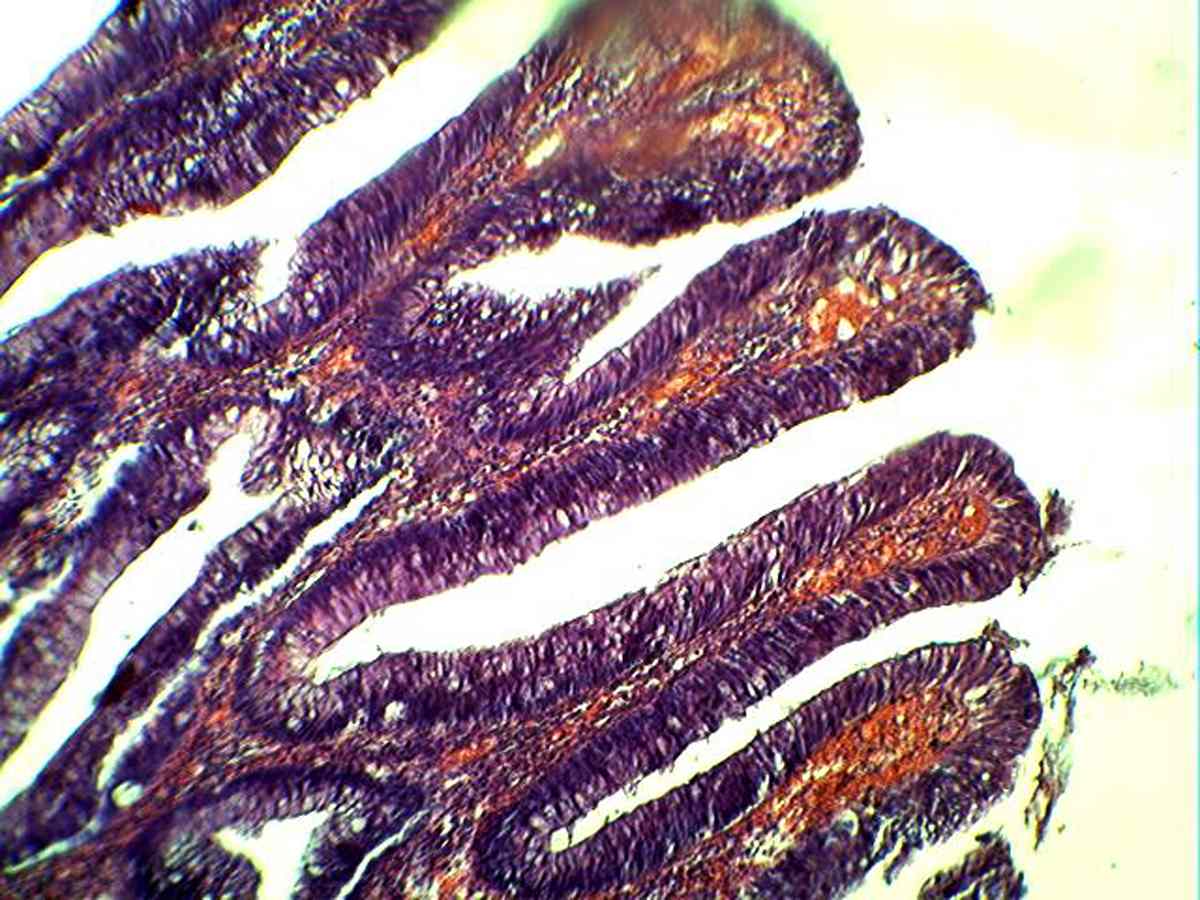
Figure 4: Villous Adenoma showing high grade dysplasia (H&E stain) (x20).
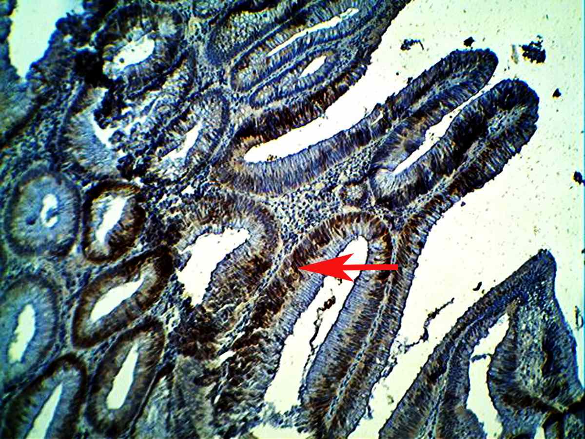
Figure 5: Villous adenoma of high grade dysplasia showing positive immunohistochemical expression of Ki-67 (Arrows) (x10).
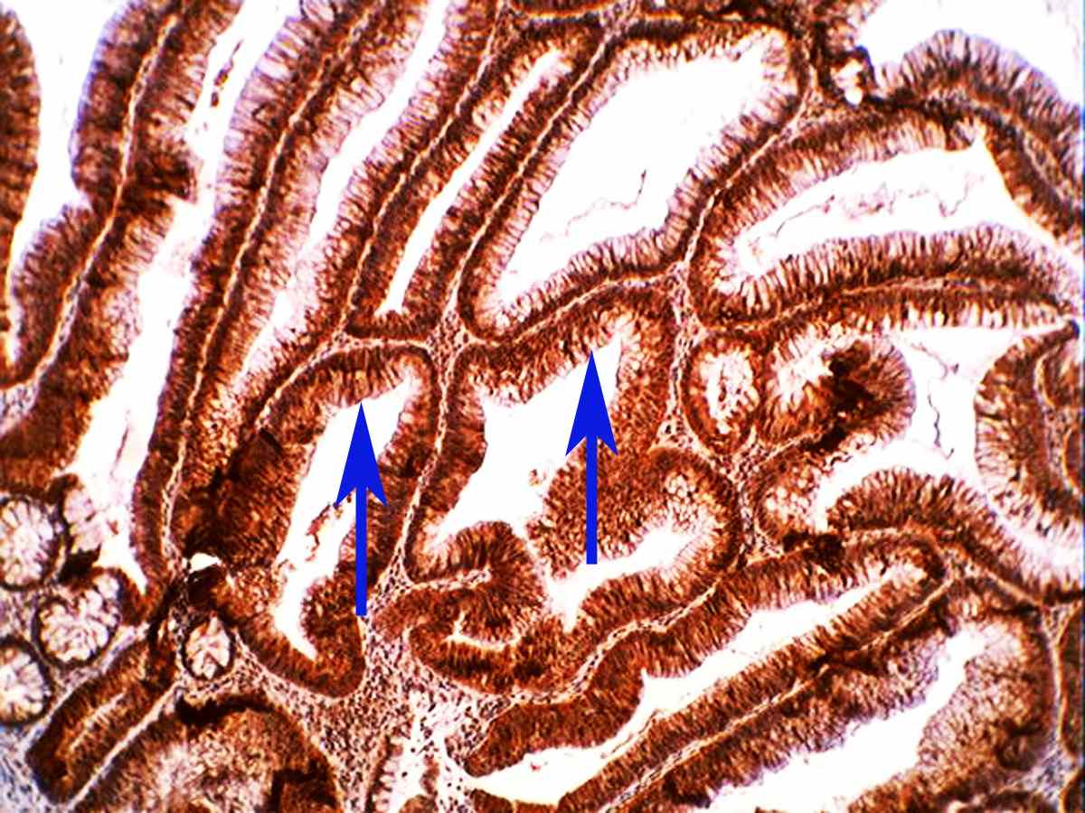
Figure 6: Villous adenoma with high grade dysplasia showing positive immunohistochemical expression of P53 (Arrows) (x20).
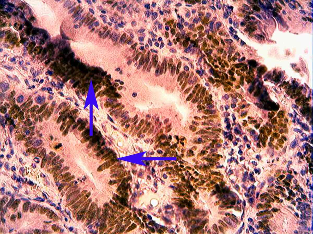
Figure 7: Tubulovillous adenoma with high grade dysplasia showing positive immunohistochemical expression of Ki-67, (arrows), (x40).
On the other hand, there was no significant correlation between the age and the expression of P53 and Ki-67 in different types of colorectal adenomas, p>0.05. R= 0.082 for p53, and for ki-67 = 0.129. (Fig. 8)
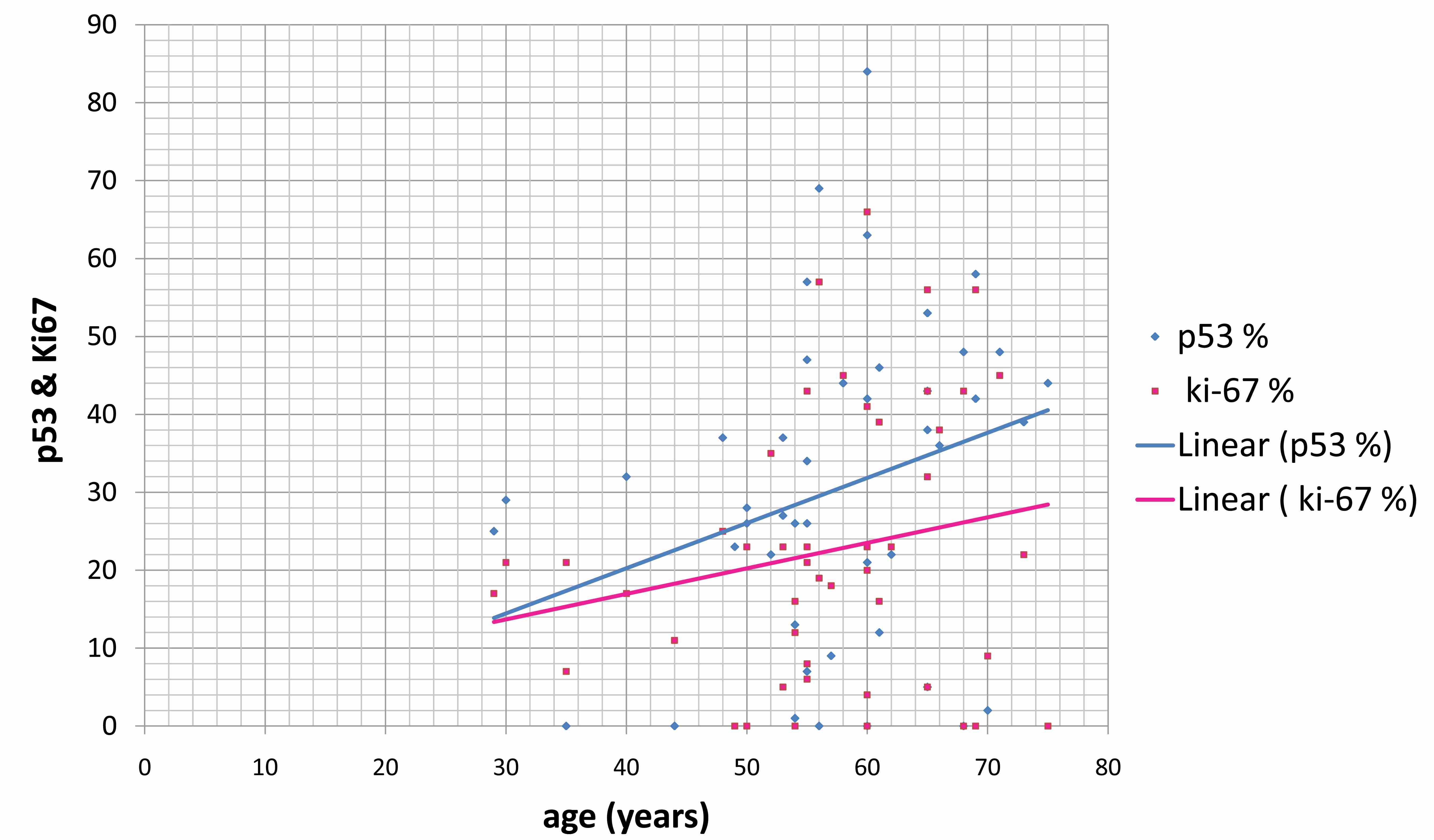
Figure 8: Effect of Age on the expressions of both Ki-67 and P53.
Also there was no significant correlation between gender and immunohistochemical expressions of both Ki-67 and P53; p>0.05. (Table 5)
Table 5: Effect of Gender on Expression of both Markers.
|
Immunohisto-chemical Markers
|
Gender
|
Number
of cases
|
Mean of expression (LI)
|
SEM
|
p value
|
|
Ki-67
|
Male
|
29
|
22.62
|
3.466
|
0.813
|
|
Female
|
18
|
21.33
|
4.022
|
|
P53
|
Male
|
29
|
27.72
|
4.339
|
0.357
|
|
Female
|
18
|
33.50
|
3.602
|
Regarding the site, also there was no significant correlation between the site of colorectal adenomas and the immunohistochemical expression of Ki-67 and P53; p>0.05. (Fig. 9)
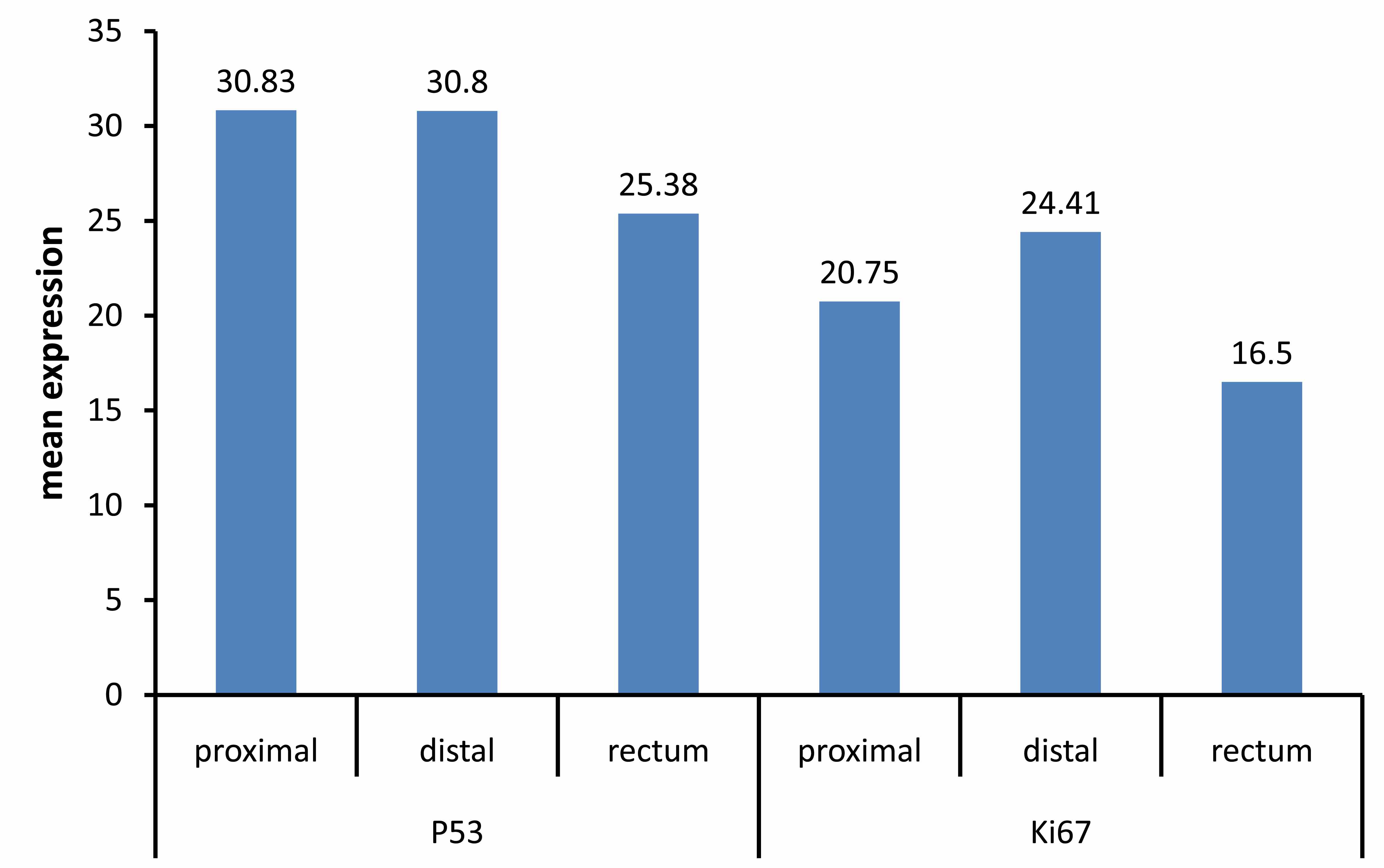
Figure 9: Distribution of Immunohistochemical expression of Ki-67 and P53 according to the Site of Adenomas in Colorectum.
Regarding the type of adenoma, there was no significant correlation between the type of adenoma and the expression of both immunohistochemical markers (P53 and Ki-67), p>0.05. (Fig. 10)
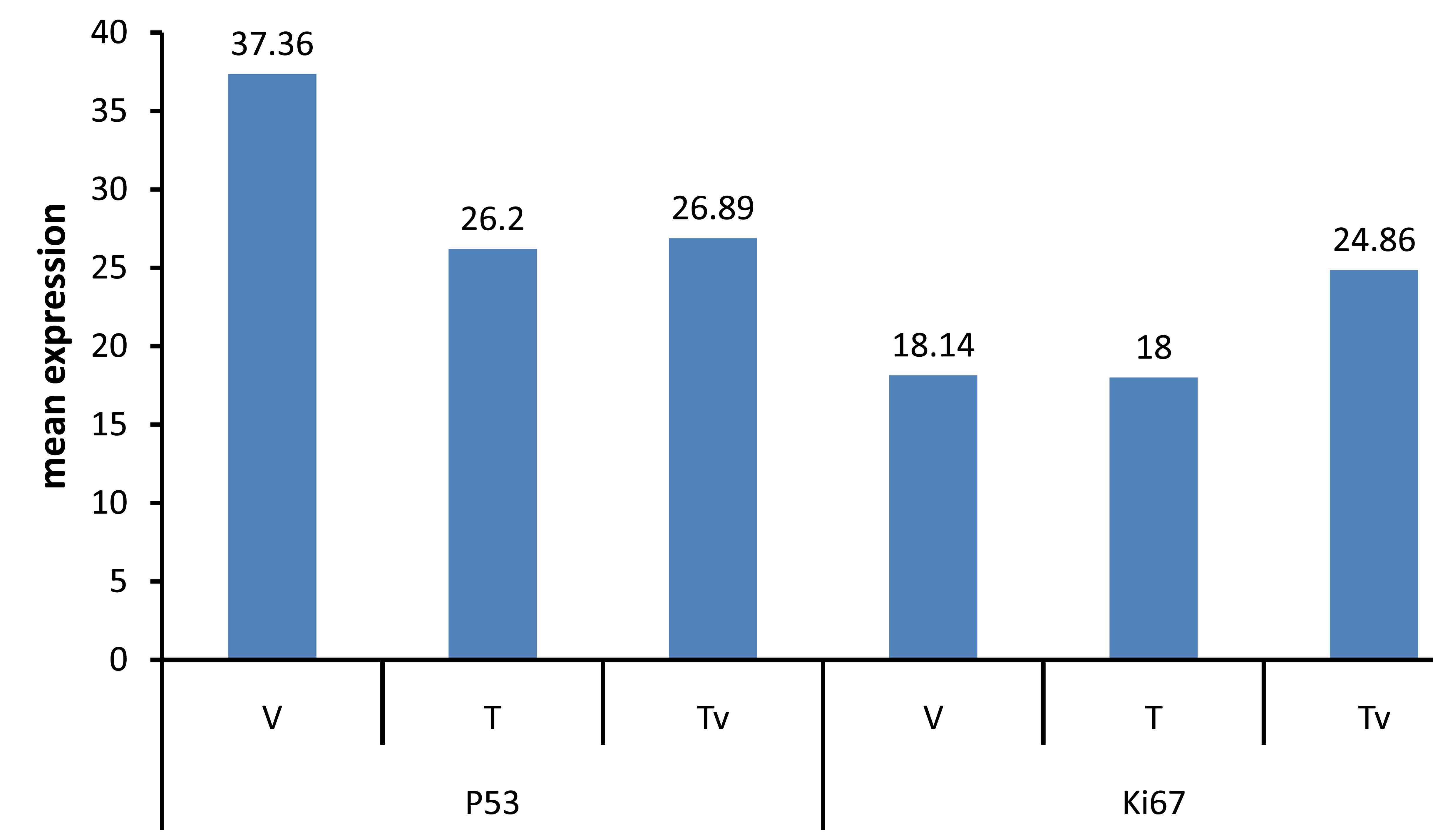
Figure 10: P53 and ki-67 Immunohistochemical expression in different Types of Colorectal Adenomas. (v= villous; t= tubular; tv= tubulovillous adenomas).
Studying the correlation of the expression of both markers, shows a positive correlation, however this correlation was not significant statistically, (p=0.130, R=0.217). (Fig. 11)
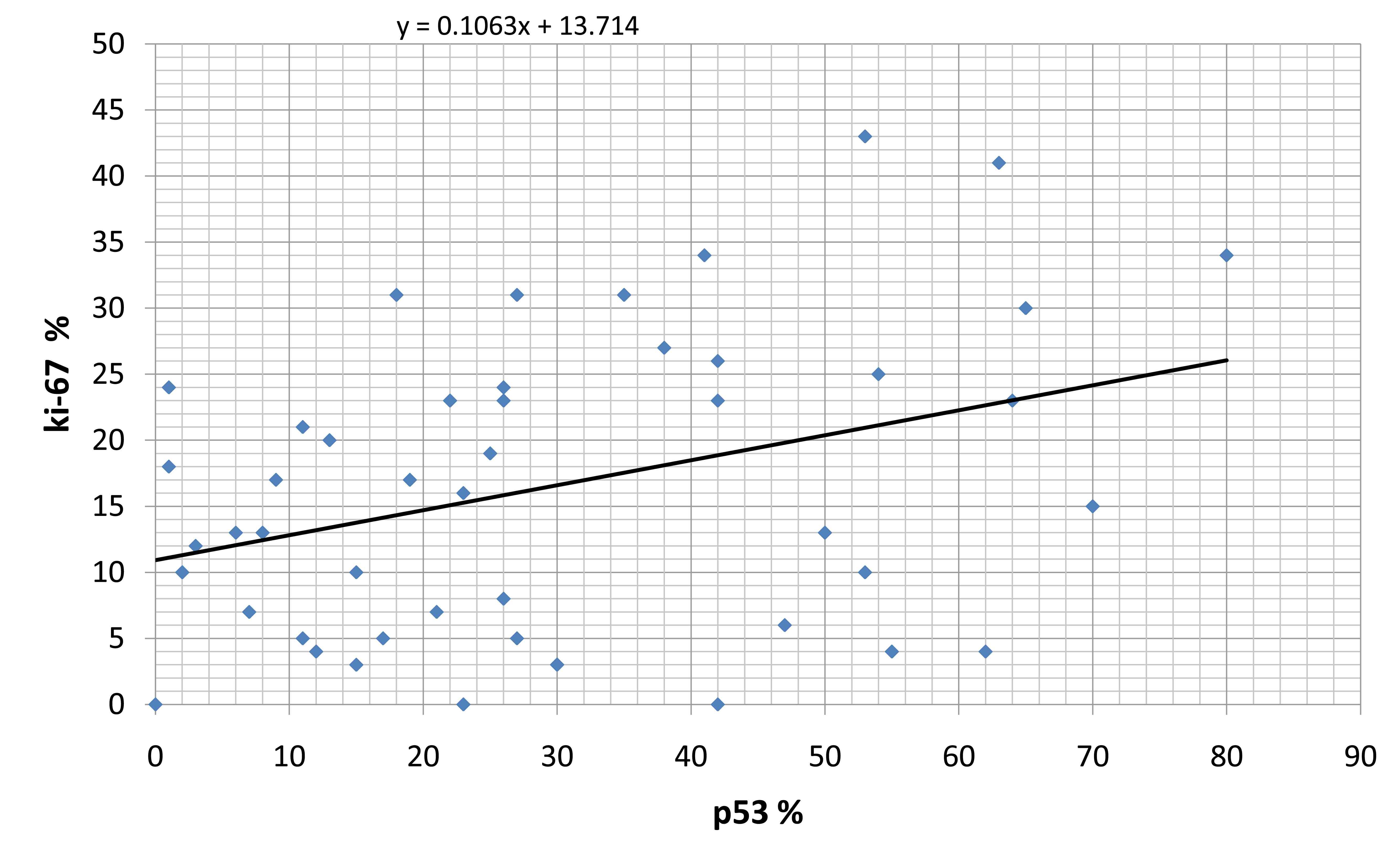
Figure 11: Correlation between Ki-67 and P53 immunohistochemical expressions in Colorectal Adenomas.
Discussion
The concept that colorectal cancers may arise from pre-existing adenomas is now widely accepted, based on epidemiological, clinical, postmortem, and molecular biological studies.13
Although this is not a large epidemiological study, it generally revealed that males constituted 59.57% of the studied patients with an age group of 50-69 years (72.34% of cases). Our data agrees with a recent study by Khatibzadeh et al. and a study by Andrey and Alexander, in the Russian Federation respectively.14,15
Considering the clinicopathological parameters; this study revealed that tubulovillous adenomas were the predominant type, followed by villous adenomas. This disagrees with previous studies by Khatibzadeh et al. in Iran; and Quirke et al. in the UK. The discrepancy may be attributed to the differences in sample size, besides the differences in racial and geographical factors, in addition to dietary habits.15,16 Although adenomas with high grade dysplasia in this study were ranked the second after low grade dysplasia; its percentage (43%) can be considered relatively high when compared with studies by Khatibzadeh et al. in Iran; Andrey and Alexander in the Russian Federation; and Quirke et al. in the UK. This can be explained sufficiently since individuals in Iraq do not undergo frequent colonoscopy, which is the most effective method of decreasing fatality of colon neoplasm, therefore are presented late and usually with high grade dysplasia.14-16
There was no significant correlation observed between the type of colorectal adenoma and the grade of dysplasia, and this result disagreed with study done by Andera et al.17
In the assessment of the relationship between Ki-67 and P53 Immunohistochemical expressions with regards to age, gender, site and type of adenoma, the present study showed that there was no significant correlation among Ki-67 and P53 expression with these parameters (p>0.05), similar findings have also been reported by Yinghao et al. in the USA.18
The present study revealed a highly significant correlation between Ki-67 and P53 expressions with the size and grade of dysplasia in colorectal adenoma, (p=0.002, 0.001); (0.006, 0.002), respectively; which is in concordance with the results obtained in Serbia by Radovanovic et al. and Abdulamir et al. in Iraq, but it disagrees with the study by Karamitopoulou et al. in Switzerland and the study by Vernillo et al. in Italy which showed that there was no significant relationship between Ki-67 expression and the grade of dysplasia. This variation is likely due to several factors, including different methods for Ki-67 and P53 analyses, besides the use of fresh frozen biopsies sometimes for these studies, population differences, selection bias for patient criteria in addition to racial and environmental factors.8,12,19,20 Although there was a generally a positive correlation between Ki-67 expression and P53 expression, it was statistically not significant (p=0.130); and this finding is in accordance with the findings of Vernillo et al. in Italy.8 In general, this study revealed that Ki-67 and P53 Immunnohistochemical expressions was significantly correlated with the size and grade of dysplasia in colorectal adenomas, however, there was no significant correlation between Ki-67 and P53 expressions with age, gender, site, and type of colorectal adenomas. The immunohistochemical expression of these markers in colorectal adenomas showed a positive correlation, however, it was not statistically significant. Thus, the immunohistochemical expression of Ki-67 and P53 may be included as part of routine pathological evaluations with other conventional prognostic factors in patients having dysplastic colorectal adenomas.
Conclusion
In summary, high grade dysplasia with significant positive immunohistochemical Ki-67 and P53 markers could be valuable parameters for selecting patients deemed to be most deserving of close surveillance in follow-up cancer prevention programs from the total adenoma population. It is closely linked with increasing age, particularly in patients with a large size adenoma of villous component in their histology.
Acknowledgements
The authors reported no conflict of interest and no funding was received for this work. |