Pindborg Tumor in an Adolescent
Kafil Akhtar,1 Nazoora Khan,2 Sufian Zaheer,2 Rana Sherwani,2 Abrar Hasan3
Akhtar K, et al. OMJ. 25, 47-48 (2010); doi:10.5001/omj.2010.12
ABSTRACT
Calcifying epithelial odontogenic tumor (Pindborg tumor), is a rare benign odontogenic neoplasm representing about 0.4-3% of all odontogenic tumors. This tumor more frequently affects adults in the age range of 20-60 years, with a peak incidence in the 5th decade of life. Calcifying epithelial odontogenic tumour has a much lower recurrence rate than ameloblastoma and malignant transformation, and metastasis is rare.
From the 1Department of Pathology, College of Medicine, K.K.U, Abha, Saudi Arabia, 2Department of Pathology, 3Department of ENT, J.N.M.C.H., A.M.U., Aligarh, INDIA.
Received: 12 Oct 2009
Accepted: 21 Nov 2009
Address correspondence and reprint requests to: Dr. Kafil Akhtar, Department of Pathology, College of Medine, KKU, Abha, Saudi Arabia.
E-mail: drkafilakhtar@gmail.com
INTRODUCTION
The calcifying epithelial odontogenic tumour (CEOT) is a rare tumour. It was first described as a separate pathologic entity by a Dutch pathologist Jens Jorgen Pindborg in 1955.1
CASE REPORT
A 16 year old male, presented with a hard nodule, 6.5 x 4.5 cm in size, on the buccal aspect of the right molar region of the mandible above the angle of the jaw. The swelling was tender and increased progressively over a period of 1 year. Roentgenogram on admission revealed a loculated /trabeculated radioluscent cyst above the angle of right mandible measuring 6 x 4 cm. The provisional clinical diagnosis of Aneurysmal bone cyst/ Ameloblastoma/ odontogenic keratocyst was made.
Resection of the right mandible from the first bicuspid through the condyle including the whole growth was performed and the specimen was submitted for histopathology.
Gross examination revealed a globular bony tissue with attached soft tissue piece measuring 6 x 3 cms. On cut section, a cystic growth was seen with bony loculations, along with cartilaginous to haemorrhagic areas. (Fig. 1)
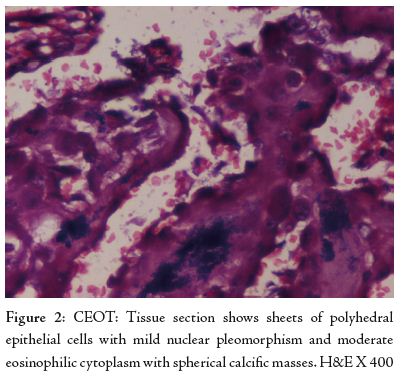
Microscopic examination of the tissue section revealed sheets of polyhedral epithelial cells in definite lobules. The closely packed cells were small with mild nuclear pleomorphism and moderate eosinophilic cytoplasm. Numerous spherical calcified masses were seen in a background of cellular degeneration with scant fibrous stroma, (Fig. 2). The diagnosis of calcifying epithelial odontogenic tumour (Pindborg tumour) was made.
DISCUSSION
CEOT is a rare, benign, but locally aggressive tumour, which accounts for less than 1% of all odontogenic tumours and most often located in the posterior mandible.2,3 Local recurrence rates of 10-15% have been reported and malignant transformation is rare.3
Etiology of this lesion is not clear. Majority of the investigators are of the opinion that, the tumour cells originate from the striatum intermedium of the normal dental lamina.4 An idea based on the morphologic similarity of the tumour cell to the normal cells of stratum intermedium and a finding of high activity of alkaline phosphatase and adenosine triphosphate at both sites.5
According to Cicconetti and colleagues, the tumor more frequently affects adults in the age range of 20-60 years, with a peak incidence in the 5th decade of life with equal sex predisposition.6 There is a marked predilection for the molar-premolar area of mandible with about 50% of cases associated with un-erupted or embedded teeth.4
Roentgenologically, this tumour is often mistaken for dentigerous cyst or ameloblastoma.4 Quite similarly; the radiographic findings in this case showed a loculated /trabeculated radioluscent cyst.
The diagnosis of calcifying epithelial odontogenic tumour is based on histological examination, revealing polyhedral neoplastic cells which have abundant eosinophilic, finely granular cytoplasm with nuclear pleomorphism and prominent nucleoli. Most of the cells are arranged in broad ramifying and anastomosing sheet-like masses with little intervening stroma; similar morphologic features were visualized in this case study. An extracellular eosinophilic homogenous material staining like amyloid is characterstic of this tumour with concentric calcified deposits, resembling psammoma bodies called “Liesegang rings.” This case also depicted calcific foci in abundance and also fused amorphous calcareous aggregates.
The CEOT is considered to have a lower recurrence rate than that of ameloblastoma and malignant transformation and metastasis is rare.3 The study patient underwent hemimandibulectomy and no recurrence was reported in 6 months of follow up.
CONCLUSION
The calcifying epithelial odontogenic tumor (CEOT), or Pindborg tumor, is a benign infiltrative odontogenic tumor that is one of the rarest. It is an infiltrative neoplasm and causes destruction with local expansion. Definitive resection of the entire mass with tumor-free surgical margins (en bloc resection) is the preferred treatment as tumor will recur if not completely removed and long-term follow ups are recommended.
ACKNOWLEDGEMENTS
The authors reported no conflict of interest and no funding has been received on this work.
-
Pindborg, J J. Calcifying epithelial odontogenic tumors. Acta Path Microbiol. Scand 956; 71:111.
-
Bridle C, Visram K, Piper K, Ali N. Maxillary calcifying epithelial odontogenic (Pindborg) tumor presenting with abnormal eye signs: case report and literature review. Oral Surg Oral Med Oral Pathol Oral Radiol Endod 2006; 102(4):12-15.
-
Ungari C, Poladas G, Giovannetti F, Carnevale C, Iannetti G. Pindborg tumor in children. J Craniofac Surg 2006 Mar; 17(2):365-369.
-
Goldenberg D, Sciubba J, Koch W, Tufano RP. Malignant odontogenic tumors: a 22-year experience. Laryngoscope 2004 Oct; 114(10):1770-1774.
-
Franklin CD, Pindborg JJ, The calcifying epithelial odontogenic tumor. A review and analysis of 113 cases. Oral Surg Oral Med Oral Pathol 1976 Dec; 42(6): 753-765.
-
Cicconetti A, Tallarico M, Bartoli A, Ripari A, Maggiani F. Calcifying epithelial odontogenic (Pindborg) tumor. A clinical case. Minerva Stomatol 2004 Jun; 53(6): 379-387.

