Isolated Fallopian Tube Torsion with Pregnancy- A Case
Report
Usha Varghese,1 Aurora Fajardo,1 T. Gomathinayagam2
Varghese U, et al. OMJ. 24, 128-130 (2009); doi:10.5001/omj.2009.27
ABSTRACT
Isolated torsion of the fallopian tube occurring in pregnancy is very rare. This entity should be considered in the differential diagnosis of acute pelvic pain with cystic adnexal mass associated with a normal ipsilateral ovary. This is a case report of a primi gravida at 29 weeks of pregnancy who was presented with isolated fallopian tube torsion.
From the 1Department of Obstetrics and Gynecology, Buraimi Hospital, Buraimi, Oman 2Department of Radiology, Buraimi Hospital, Buraimi, Oman.
Reviewed: 19 Dec 2008
Accepted: 30 Jan 2009
Address correspondence and reprint requests to: Dr. Usha Varghese, Department of Obstetrics and Gynecology, Buraimi Hospital, Buraimi, Oman
E-mail: umt24@yahoo.comINTRODUCTION
Torsion of the fallopian tube without ovarian torsion is a rare, but noteworthy, cause of lower abdominal pain in women of reproductive age. Adnexal torsion can occur in all ages. Expedient diagnosis is important to prevent adverse sequelae. Since the symptoms are non specific, diagnosis can be challenging. This is a case report of isolated fallopian tube torsion with pregnancy from Buraimi Hospital, Oman.
CASE REPORT
A 26 year old Omani woman primigravida, was admitted to the delivery suite of Buraimi Hospital, Oman, at 29 weeks gestation with complaints of acute abdominal pain since the previous day. The patient experienced pain which was associated with nausea. The patient had no history of fever.
Physical examination showed that the patient was afebrile and vital signs were stable. The abdomen was soft with fundal height corresponding to the period of gestation. The uterus was relaxed with a regular fetal heart. No uterine contractions were noted. Tenderness was present in the left lower abdomen. The vaginal examination showed a closed cervix without any evidence of bleeding or any abnormal discharge.
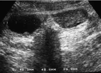
An ultrasonogram revealed a single live foetus in cephalic presentation. Parameters were corresponding to 29 weeks gestation. Estimated fetal weight was 1.500 kg. The placenta was seen on the right lateral uterine wall. There was more liqour than normal for the gestational age. There were suggestions of two ovoid masses (42.3 and 48.8 mm) with a thin hypoechoic “bridge” between them seen at the left of the uterus and measuring about 89.9 mm in total length. The superomedial mass was anechoic with a small echoic focus posteriorly while the inferolaterally situated mass was hypoechoic with uniform low level echoes within it. They had an appearance of a single bilobed structure. Both showed evidence of good posterior enhancement (figure 1). There was no vascular flow on Doppler examination. Both ovaries were not visualized. In view of the above features the possibilities of (a) left ovarian cyst and (b) Left adnexal torsion were considered.
Laboratory parameters were as follows; Hemoglobin 10.4 g/dl, Hematocrit (HCT) 30.6%, Platelets-211 k/ul, wbc-8.8k/ul, Gr-76.5%, Lymph 27.2%, Mid-5.3%, Blood Group-B+, Sickling-Negetive, Veneral Disease Research Laboratory (VDRL) Non Reactive, Urine analysis-Normal.

Figure1: Both showed evidence of good posterior enhancement.
In view of acute abdominal pain with possible left adnexal torsion, the patient was taken up for emergency exploratory laparotomy after obtaining informed consent. Intraoperative findings were as follows: there was a twisted dusky haemorrhagic mass of 6 x 6 cm at the left fallopian tube involving the fimbriae. There was haemoperitoneum and old dark blood was seen at the left pelvic side wall. The left ovary looked healthy. This was proceeded with left salpingectomy, as the left fallopian tube was not salvageable. The right ovary and right fallopian tube appeared healthy. She was put on intravenous antibiotics and received 2 doses of betamethasone injection. There was no indication of any preterm labour. The postoperative period was uneventful and the patient was discharged on the fifth postoperative day in good condition. The patient was advised regular follow up at the antenatal clinic. The histopathology report showed torsion of the left fallopian tube with haematosalpinx.
DISCUSSION
Isolated torsion of the fallopian tube is a rare but noteworthy cause of lower abdominal pain in women of reproductive age.1 It is a difficult condition to evaluate clinically and surgery is often necessary to establish the diagnosis.2 It occurs without ipsilateral ovarian involvement associated with pregnancy, haemosalpinx, hydrosalpinx, ovarian or par ovarian cysts and other adnexal alteration or even with an otherwise normal fallopian tube.3 The exact cause of fallopian tube torsion is unknown.4 Youssef et al noted factors that could possibly influence the occurrence of fallopian tube torsion and divided them into two categories; Intrinsic factors such as congenital anomalies of the fallopian tube, acquired pathology of the fallopian tube for example; hydrosalpinx, hematosalpinx, neoplasm, surgery, autonomic dysfunction and abnormal peristalsis, and Extrinsic factors for example, changes in the neighbouring organs such as neoplasm, adhesions, pregnancy, mechanical factors, movement or trauma to the pelvic organs, or pelvic congestion.5
Collectively, the existing reports indicate that the mechanism underlying tubal torsion is apparently a sequential mechanical event. The process begins with the mechanical blockage of the adnexal veins and lymphatic vessels by ovarian tumour, pregnancy, hydrosalpinx or pelvic adhesions after tubal infection or pelvic operation. This obstruction causes pelvic congestion and local edema with subsequent enlargement of the adnexa, which in turn induces partial or complete torsion.6 The incidence of fallopian tube torsion occurs in 1/1.5 million women. Sporadic cases are reported each year.4 The right fallopian tube is commonly affected. This may be due to the presence of the sigmoid colon on the left side to slow venous drainage on the right side which may result in congestion.7,8 But in this case report, it was the left tube which was affected.
Presenting symptoms for tubal torsion include sudden onset of lower quadrant pain that can be constant and dull or paroxysmal and sharp radiating to the thigh or groin, nausea and occasional fever.9 A tender adenexal mass may be palpable. Ultrasonographic findings show that the ipsilateral ovary appeared normal, free fluid, a dilated tube with thickened echogenic walls and internal debris or a convoluted echogenic mass.10, 11 High impedence, reversal or absence of vascular flow in the tube has also been reported although, in practice, confident spectral Doppler analysis of the tubal wall may be difficult.12 Reported Computed Technology (CT) findings of isolated tubal torsion include an adnexal mass, a twisted appearance to the fallopian tube, dilated tube greater than 15mm, a thickened and enhancing tubal wall and luminal CT attenuation greater than 50HU consistent with haemorrhage. Secondary signs include free intrapelvic fluid, peritubular fat stranding, enhancement and thickening of the broad ligament and regional ileus.10,13 The available laboratory or imaging studies cannot confirm fallopian tube torsion.4 Ultimately the diagnosis is generally made at the time of surgical exploration. Prompt consideration of this diagnosis and surgical detorsion may prevent irreversible vascular changes. Complications from tubal torsion include fallopian tube necrosis and gangrenous transformation leading to peritonitis.9
Differential diagnosis of adnexal torsion should be considered in any girl or woman with lower abdominal pain and adnexal mass. Other diagnostic possibilities include appendicitis, renal colic, urinary tract infection, regenerating leiomyoma, abruption of the placenta, hemorrhagic or ruptured ovarian cyst etc.
Laparoscopy is currently the most specific diagnostic tool for evaluating torsion and is used for the treatment of this condition. Currently, laparoscopic adnexal detorsion, not adnexectomy is the procedure of choice.4 This is because most of the patients are in their reproductive years, efforts should be made to preserve fertility if the ischaemic damage appears to be reversible, and no malignancy is suspected. A complete resection is performed when the tissue is gangrenous, or if there is tubal or ovarian neoplasm or the woman has completed her family. When there is no apparent ischemic damage, most of the twisted adnexa regain their function.
Recovery is much faster after laparoscopy than laparotomy. Laparoscopy also causes fewer pelvic adhesions which is particularly important for women of reproductive age who wish to preserve their fertility. If the patient is in her third trimester, most surgeons prefer laparotomy, because laparoscopy is technically very difficult.4 In this case, the patient was in her third trimester with gangrenous left fallopian tube and hence underwent laparotomy and left salpingectomy.
CONCLUSION
Isolated fallopian tube torsion is a rare entity. Adnexal torsion should be considered in any girl or woman with sudden onset of lower abdominal pain with an adnexal mass. Imaging techniques may be suggestive but not conclusive. Expedient diagnosis is important to prevent adverse sequelae. However, the diagnosis can be challenging because the symptoms are relatively non specific.
REFERENCES
1. Batukan C, Ozgun MT, Turkyilmaz C,Tayyar M. Isolated torsion of the fallopian tube during pregnancy : A case report. J Reprod Med 2007; 52:745-747.
2. Lineberry TD, Rodriguez H. Isolated torsion of the fallopian tube in an adolescent.a case report. J paed Adolesc Gynae 2000; 13:135-137.
3. Antoniou N, Varras M, Akrivis C, Kitsiou E, Stefanaki S, Salamalakis E. Clin Expt Obstet Gynecol 2004; 31:235-238.
4. Krissi H, Shalev J, Bar-Hava I, Langer R, Herman A, Kaplan B. Fallopian tube torsion: Laparoscopic evaluation and treatment of a rare Gynecological entity. J Am Board of Fam Pract 2001; 14:274.
5. Youssef AF, Fayad MM, Shafeek MA.Torsion of the fallopian tube. A clinico-pathological study. Acta Obstet Gynec Scand 1962; 41:292-309.
6. Bernardus RE, Van der Slikke JW, Roex AJ, Dijkhuizen GH, Stolk JG. Torsion of the fallopian tube: Some considerations on its etiology. Obstet Gynecol 1984; 64:675-678.
7. Phupong V, Intharasakda P. Twisted fallopian tube in Pregnancy, a case report. BMC Pregnancy and childbirth. 2001; 1:5.
8. Yalcin OT, Hassa H, Zeytinoglu S, Isiksoy S. Isolated torsion of fallopian tube during pregnancy; Report of two cases. Eur J Obstet Gynecol Reprod Biol. 1997; 74:179-182.
9. Ferrera PC, Kass LE, Verdile V. Torsion of the fallopian tube. Am J Emerg Med 1995; 13:312-314.
10. Ghossain MA, Buy JN, Bazot M , Haddad S, Guinet C, Malbec L, et al. CT in adnexal torsion with emphasis on tubal findings:correlation with US. J Comput Assist Tomogr 1994; 18:619-662.
11. Propeck PA, Scanlan KA. Isolated fallopian tube torsion. AJR 1998; 170:1112-1113.
12. Baumgartel PB, Fleischer AC, Cullinan JA, Bluth RF. Color Doppler Sonography of tubal torsion. Ultrasound Obstet Gynecol 1996; 7:367-370.
13. Hiller N, Baum LA, Simanovsky N, Levsagi A. Aharoni D, Sella T. Am J Roentgenol 2007; 189:124-129.