|
Abstract
This is a case of a 17-year-old boy who was admitted to the clinic suffering from right hip-inguinal region pain which began after a soccer match. He had an avulsion fracture in the anterior inferior iliac spine and CT/MR images showed similarities to a bone tumor. These fractures are especially seen in the apophyses which are within the ossification process. The radiological appearance of these fractures during the healing period may be involved with bone tumors. In the control MRI of the patient after one year, the fracture was almost healed completely. Treatment is generally conservative. Good results have been reported with rest, analgesic and anti-inflammatory drugs. Familiarity with radiological findings of these avulsion fractures in this region not only protects the patient from invasive procedures but also avoids the involvement of the practitioner in legal consequences resulting from a misdiagnosis of sarcoma.
Keywords: Avulsion fracture; Pelvic apophyses; MRI.
Introduction
Acute avulsion fractures of the pelvic apophyses are uncommon traumas. This clinical condition is usually encountered in adolescents, who engage in physical activities such as ball kicking, running and jumping. During such physical activities, avulsion fracture of the pelvic bone can occur due to an abrupt and strong traction to the pelvic spine by the musculotendinous junction of the muscles. These fractures are seen especially in the apophyses which are within the ossification process.1 The radiological appearance of these fractures during the healing period may be confused with bone tumors.2 The aim of this report is to emphasize apophysial fracture of the anterior inferior iliac spine for awareness of this injury, appropriate interpretation of imaging findings and for avoidance of unnecessary biopsy.
Case Report
A 17-year-old boy was admitted to a health clinic about nine months ago, suffering from right hip-inguinal region pain which began after a soccer match. A soft tissue injury was diagnosed and palliative treatment was started. He was then admitted to the clinic due to recurrent pain. The patient stated that he had continued to play soccer during this period. On physical examination, there was tenderness and swelling in the right inferior iliac region. Pain increased with motion in the right hip and pelvic region. Cortical irregularity at the inferior margin of the right iliac bone and ossification within the adjacent soft tissues were seen in the pelvic radiographs. Decision was made to perform CT and MR imaging to make further evaluations and to ascertain whether or not there was an underlying pathological fracture. The CT exam revealed a fracture line with irregular margins at the anterior inferior iliac bone, ossification through the adjacent soft tissues and a suspicious lytic lesion which was expanding the cortex. (Fig. 1)
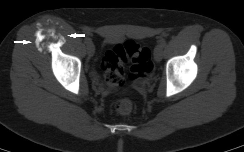
Figure 1: Axial CT image showing marked irregularity and the expansion of the bone in the anterior right iliac and the superior acetabular region.
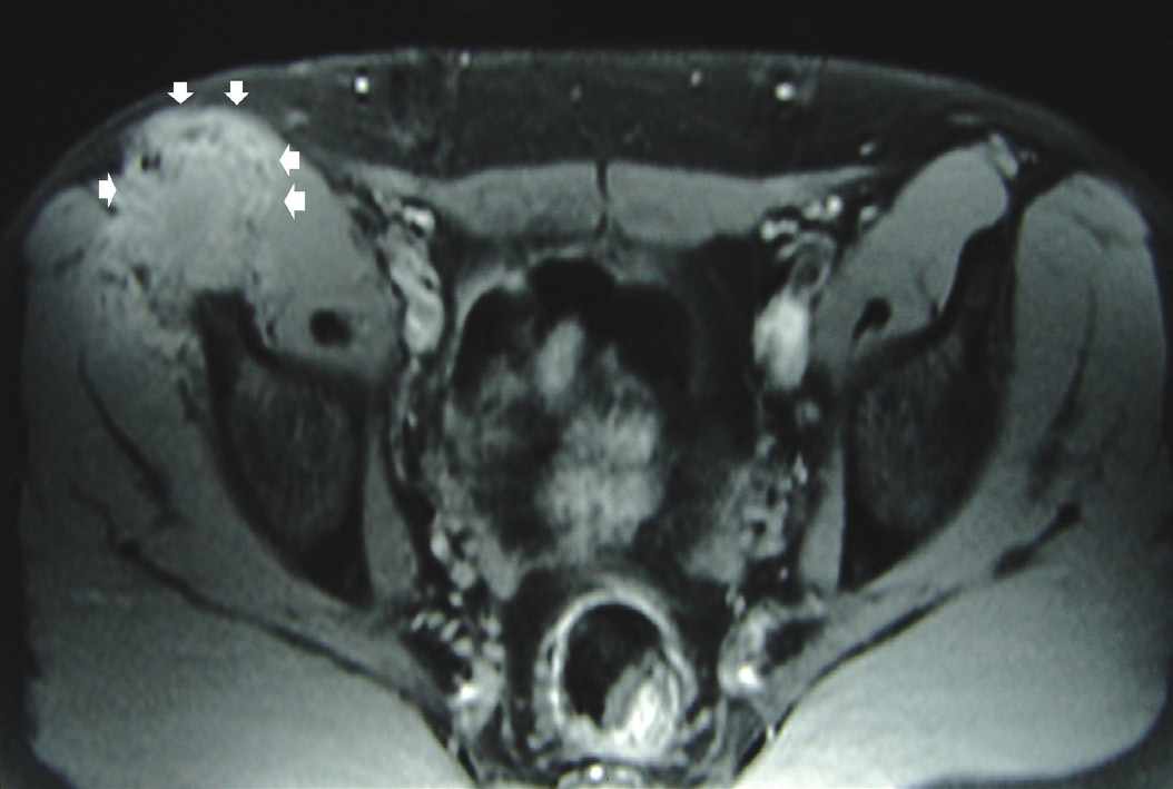
Figure 2: Axial T1W pre-contrast image showing damage to the anterior right iliac bone and the adjacent soft tissue. Note the high signal intensity areas suggesting hematomas around the soft tissue margins (arrow heads).
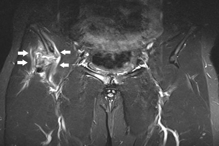
Figure 3: Fat-saturated T2W coronal image showing the transverse fracture line in the right iliac bone with irregular margins and expansion, edema and hemorrhage areas including callus formation in the adjacent soft tissues (arrows).
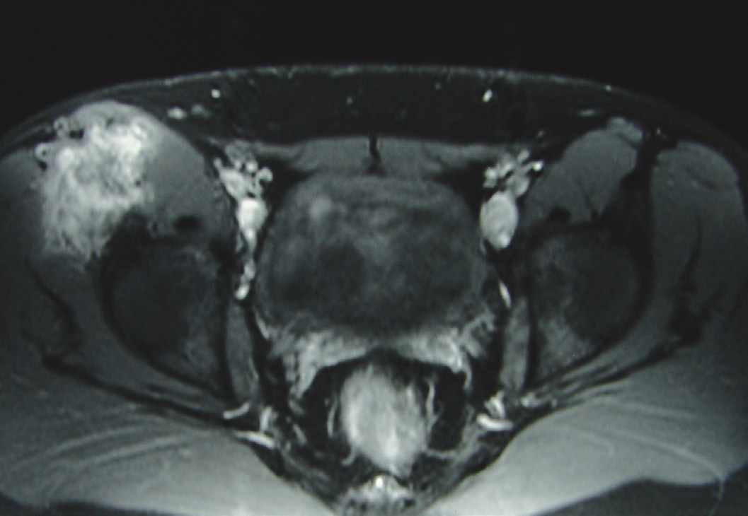
Figure 4: Axial post-intravenous gadolinium T1W fat-saturated image showing marked enhancement through the fracture line and in the adjacent soft tissues. It is mimicking a bone tumor with this aggressive appearance.
The fracture line with irregular margins was also seen in the MR images (Figs. 2 and 3). After intravenous contrast injection, significant enhancement through the soft tissues was evident (Fig. 4). There was increased signal intensity consistent with edema at the rectus femoris muscle and adjacent soft tissues, especially in the fat-saturated T2W images. Although a biopsy through the enhanced soft tissues was considered to rule out any possible underlying malignancy, the treatment recommended was to rest and not play soccer. The final decision on the patient was based on the MR images because of the possible risk of confusion of histopathological diagnosis with healing fractures and sarcomatous lesions. At the one year follow-up examination, his clinical status had improved, and the tenderness and swelling in the iliac region had disappeared.
On MR imaging, the fracture was seen to have healed almost completely and there was no soft tissue component (Fig. 5). Therefore, it was decided that this fracture, which was seen in an adolescent, was an avulsion fracture and there was no need to consider it as pathological.
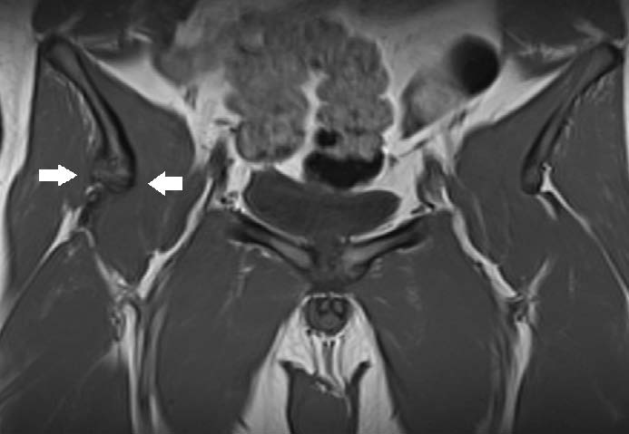
Figure 5: Coronal T1W image at 12-month follow-up showing the fracture in the right iliac bone has almost healed completely and the associated soft tissue component has disappeared (arrows).
Discussion
Secondary ossification centers located on the ends of the long bones are named epiphyses and those at the insertion site of a muscle are named apophyses. Ossification of the pelvic bone apophyses occurs during adolescence, which is a time of increased physical activity for most people. The ratio of muscular strength to physeal strength is greatest during early puberty when the chondrocalcinosis is not yet complete.1 The rectus femoris muscle is the most commonly injured muscle within the quadriceps muscle group because it extends between two joints (hip and knee) and is most vulnerable during kicking games such as soccer.3,4 Sports related injuries are considered to be responsible for the majority of lower limb fractures.5 Avulsion fractures of the anterior inferior iliac spine occur due to a straight pull of the head of the rectus femoris muscle,3,6 and generally occur when there is hyperextension of the hip joint and the flexion of the knee, as in the action of kicking a ball. In this condition, which is when there is maximum effect on the tendon of the rectus femoris muscle, avulsion occurs at the insertion region of the muscle in the adolescents whose apophyses are already open.1,6,7 Fractures of the anterior inferior iliac spine are more rare than fractures of the anterior superior iliac spine. As the fusion of the anterior superior iliac spine apophysis occurs later than the fusion of the anterior inferior iliac spine, the fractures of the anterior superior iliac spine are more common in older adolescents and young adults.1,7
Clinical diagnosis is made by physical examination, direct radiographs and CT or MRI when necessary.3 It is possible to make an easy and a correct diagnosis immediately after injury; however, if some time has passed and callus has formed, definitive diagnosis may be difficult because the radiological findings can be confused with malignancy.6 In the current case, the delay in diagnosis of the avulsion fracture of the anterior inferior iliac spine and the repeated trauma from playing soccer during the intervening period delayed the healing of the fracture and made the diagnosis difficult. Treatment for these fractures is generally conservative and good results have been reported with rest, analgesic and anti-inflammatory drugs.2,8 Surgical treatment can be planned when it is necessary to speed up the healing process or when there is a displaced fragment.2
Avulsion fractures with callus formation during the healing period may be confused with osteosarcoma or Ewing’s sarcoma.2,9 Dhinsa et al. reported that subtle bone marrow edema in the anterior iliac spine and hematoma (hyperintense in T1W and T2W images) suggests a fracture rather than a tumor.2
Conclusion
Adolescents are an age group who engage in intense physical activity and are most likely to experience apophysial injuries. There are very few reports in the literature of MRI findings of avulsion fractures of the direct head of the rectus femoris muscle.2,3 While it is very easy to diagnose these avulsion fractures in this region with direct radiographs, MRI findings may be very confusing especially in cases where there is callus formation during the healing period. Being familiar with clinical management and the radiological findings of these avulsion fractures in this region not only prevents the patient from having to undergo invasive procedures such as biopsy but also allows the practitioner to avoid legal consequences which may result from a misdiagnosis of sarcoma.
Acknowledgements
The authors declare that no benefits or funds were received in support of this study.
References
1. Atalar H, Kayaoğlu E, Yavuz OY, Selek H, Uraş I. Avulsion fracture of the anterior inferior iliac spine. Ulus Travma Acil Cerrahi Derg 2007 Oct;13(4):322-325.
2. Dhinsa BS, Jalgaonkar A, Mann B, Butt S, Pollock R. Avulsion fracture of the anterior superior iliac spine: misdiagnosis of a bone tumour. J Orthop Traumatol 2011 Sep;12(3):173-176.
3. Ouellette H, Thomas BJ, Nelson E, Torriani M. MR imaging of rectus femoris origin injuries. Skeletal Radiol 2006 Sep;35(9):665-672.
4. Bordalo-Rodrigues M, Rosenberg ZS. MR imaging of the proximal rectus femoris musculotendinous unit. Magn Reson Imaging Clin N Am 2005 Nov;13(4):717-725.
5. Dhar D, Varghese T. Audit of inpatient management and outcome of limb fractures in children. Oman Med J 2011 Mar;26(2):131-135.
6. Rajasekhar C, Kumar KS, Bhamra MS. Avulsion fractures of the anterior inferior iliac spine: the case for surgical intervention. Int Orthop 2001;24(6):364-365.
7. Gomez JE. Bilateral anterior inferior iliac spine avulsion fractures. Med Sci Sports Exerc 1996 Feb;28(2):161-164.
8. Yildiz C, Yildiz Y, Ozdemir MT, Green D, Aydin T. Sequential avulsion of the anterior inferior iliac spine in an adolescent long jumper. Br J Sports Med 2005 Jul;39(7):e31.
9. Karakas HM, Alicioglu B, Erdem G. Bilateral anterior inferior iliac spine avulsion in an adolescent soccer player: a typical imitator of malignant bone lesions. South Med J 2009 Jul;102(7):758-760.
|