|
Introduction
A 30-year-old patient was referred to our Hepatology clinic because he was incidentally found to be positive for HBsAg. For the last 18 months prior to the date of referral, he had been treated as having IBS in another hospital due to complaints of recurrent abdominal pain, abdominal bloating, and diarrhea alternating with constipation. Despite having good appetite, he had lost some weight (4 kg in 6 months). He gave no history of gastrointestinal bleeding. Also, he had no extra-intestinal symptoms or past medical history of note.
Physical examination revealed no abnormalities. Laboratory data were as follows: Total bilirubin: 15 μmol/L (3-17 μmol/L), ALT: 30 IU/L (3-35 IU/L), AST: 28 IU/L (3-35 IU/L), ALP: 200 IU/L (30-35 IU/L), albumin: 30 g/L (35-50 g/L); HBV-DNA: 50 IU/mL, HBeAg: -ve, HBe-Ab: +ve; Hb: 9.3 gm/dL, WBC: 4000/cmm, and platlets: 300,000/cmm. Peripheral blood film is shown in Fig. 1. INR was 1.6 (0.9-1.3) and TFTs revealed high TSH with normal T3 and T4. RFTs and electrolytes were normal except for hypocalcemia. Abdominal ultrasonography and CT scan with IV contrast were normal. Upper GI endoscopy was normal; however, a biopsy of the small bowel was obtained and the result of histopathological study is shown in Fig. 2.

Figure 1: Peripheral blood smear.
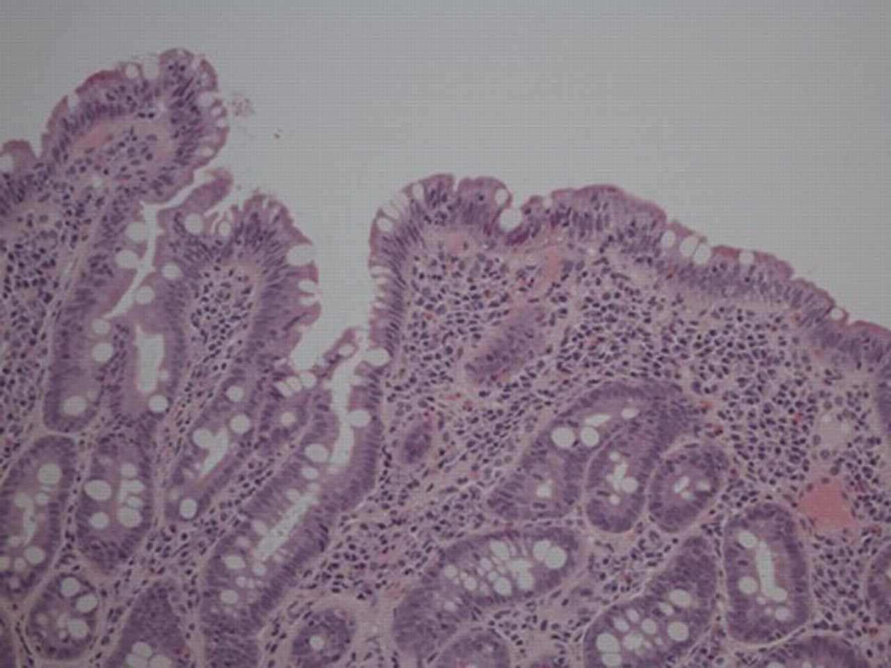
Figure 2: Histological study of small bowel biopsy.
Question
1. What is the most likely diagnosis?
2. Mention two further laboratory tests to support the diagnosis in this patient.
|