A 24-year-old married woman presented to the outpatient clinic with abdominal pain and weight and appetite loss lasting a few months. The abdomen appeared soft on clinical examination, with a palpable midline mass eliciting fluid thrill, corresponding to 32 weeks gestation. Speculum examination revealed a retroverted uterus, healthy cervix, and posterior fornix of the vagina with minimal mucous discharge. On examination of the anterior fornix of the vagina, a cystic mass was seen compressing the bladder and the structures below.
Abdominal ultrasound revealed a large cystic lesion in the midline with multiple internal echoes, measuring 20.1 × 11.5 × 13.1 cm in the abdominopelvic region. The lesion seemed to arise from the left ovary as the left ovary could not be visualized separately. On magnetic resonance imaging (MRI), a large abdominopelvic cystic mass seemed to arise from the left ovary with multiple thin septations, measuring 23.0 × 9.0 × 18.0 cm noted in the midline, displacing and compressing the adjacent structures. The classical stained glass appearance of the large cystic lesion was non-satisfactorily visualized on short tau inversion recovery images.
Questions
- What is the likely diagnosis?
- What are the differentials?
- What is stained glass appearance?
Answers
- Owing to the history and the specific findings of the cyst, which showed septations, and considering a large cystic lesion, a mucinous type of cystadenoma was suspected.
- Differential diagnoses like epithelial tumors of the ovary are considered, in which serous and mucinous cystadenomas are the most common. The others are endometrioid, clear cell, and Brenner tumors.
- A mixed-signal intensity of the lesion on T1- and T2-MRI images appears as a stained glass appearance [Figure 1 and 2].
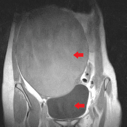
Figure 1: MRI image of the cystic lesion (red arrows) on T1-cor image.
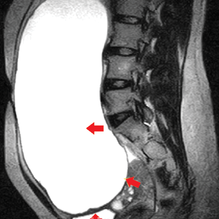
Figure 2: MRI image of the cystic lesion (red arrows) on T2 sagittal image.
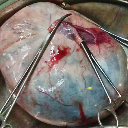
Figure 3: Gross specimen image of the cystic lesion.
Discussion
Mucinous cystadenomas are benign epithelial mucin containing tumors, often larger than serous cystadenomas and present in the reproductive age group after 20 years. Epithelial mucinous tumors have the following common characteristics: they appear multilocular both macroscopically and in images, and are large, often exceeding 10 cm.1 Depending on the histological degree of malignancy, epithelial mucinous tumors may be benign cystadenomas (85%), borderline cystadenomas (10%), or mucinous cystadenomas (5%).2
Mucinous cystadenomas have thin regular walls and several septations. They lack solid components and usually never enhance post-contrast. The cystic loculi may have variable signal intensity on both T1- and T2-weighted images, namely stained glass appearance, based on different mucin concentrations [Figures 1 and 2].3 Differentiating mucinous from serous, the latter follows the pattern of a simple cyst or multiseptated mass, whereas the former is typically a large multilocular lesion with locks of various fluid contents.4
In our case, the patient presented with abdominal symptoms. On ultrasound, a large cystic lesion measuring 20.1 × 11.5 × 13.1 cm was seen with thin internal septations and internal echoes, seen probably arising from the left ovary/adnexa, extending up to the epigastric region. On MRI, the left ovary was not seen separately. The stained glass appearance was partially appreciated [Figure 1 and 2]. A cystic neoplasm of the left ovary was to be considered. Since conservative surgery as ovarian cystectomy and salpingo-oophorectomy is adequate for benign lesions,5 she underwent left salphingo-oopherectomy laparoscopically.
Intraoperative findings showed a large cyst found replacing the left ovary. On microscopic examination, ovarian stroma with cystic spaces lined by an attenuated single layer of tall non-ciliated columnar epithelium, with apical mucin and basal nuclei (mucinous lining) was seen. On gross examination, the cystic mass measured 23.0 × 9.0 × 18.0 cm, which weighed 4.5 kg containing 4.0 L of hemorrhagic fluid. The walls of the cyst showed a smooth inner surface with multi-loculations [Figure 3].
Conclusion
Mucinous cystadenomas of the ovary are benign epithelial tumors more common at the reproductive age group and are often large cystic lesions with multi-loculations, containing mucin, which shows the classical stained glass appearance.
references
- Laurent P-E, Thomassin-Piana J, Jalaguier-Coudray A. Mucin-producing tumors of the ovary: MR imaging appearance. Diagn Interv Imaging 2015 Nov;96(11):1125-1132.
- Seidman JD, Kurman RJ, Ronnett BM. Primary and metastatic mucinous adenocarcinomas in the ovaries: incidence in routine practice with a new approach to improve intraoperative diagnosis. Am J Surg Pathol 2003 Jul;27(7):985-993.
- Foti PV, Attinà G, Spadola S, Caltabiano R, Farina R, Palmucci S, et al. MR imaging of ovarian masses: classification and differential diagnosis. Insights Imaging 2016 Feb;7(1):21-41.
- Jung SE, Lee JM, Rha SE, Byun JY, Jung JI, Hahn ST. CT and MRI of ovarian tumors with emphasis on the differential diagnosis. Radiographics 2002;22(6):1305-1325.
- Alobaid AS. Mucinous cystadenoma of the ovary in a 12-year-old girl. Saudi Med J 2008 Jan;29(1):126-128.