A 60-year-old man came to the orthopedics department with swelling of the entire right foot with multiple painful nodules, which oozed pus and blood intermittently for 25 years [Figure 1]. This caused disability and daily limitations to his lifestyle. He had presented with this complaint several times and was referred to a tertiary care center. Examination of the foot showed a swollen foot and ankle with multiple subcutaneous nodules and draining sinuses. On palpation, the nodules were tender and boggy with restriction of movements. Plain radiograph of the foot showed multiple lytic lesions in all the tarsals, metatarsals, and proximal phalanges [Figure 2]. T2-weighted magnetic resonance imaging (MRI) showed multiple lesions with classic ‘dot in circle’ sign in the bones and soft tissue [Figure 3].
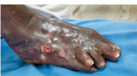
Figure 1: Swollen, oedematous foot with multiple oozing sinuses.

Figure 2: X-ray of the foot showing multiple lytic lesions seen in tarsals, metatarsals, and phalanges.
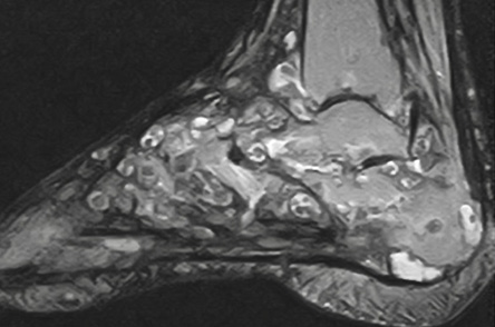
Figure 3: Magnetic resonance imaging of the foot showing multiple ‘dot in circle’ sign.
Questions
1. What is the diagnosis?
2. What is the ‘dot in circle’ sign, and what are they referring to?
3. What is the best modality for identification?
Answers
1. Mycetoma foot.
2. ‘Dot in circle’ is a classic sign in mycetoma foot. The central low signal dot represents the fungal grains, and the surrounding T2 hyperintense granulation tissue with peripheral T2 hypointense fibrous septa represents the circle.
3. Though plain X-ray shows multiple lytic lesions, MRI is the modality of choice to view the ‘dot in circle’ sign.
Discussion
Mycetoma is a chronic granulomatous disease, which is prevalent in dry tropic areas where there is low rainfall. This disease was first reported in the Madurai district of South India, hence it is named Madura foot.1 The organism that affects is a fungus or actinomycetes (a gram-positive bacilli).2 It commonly affects the feet, which are more prone to trauma and resultant infection compared to the other organs in the body such as the lower legs, hands, chest, shoulders, and arms.3 Infection occurs by direct implantation of organisms that are normal inhabitants of the soil.4 The route of entry of the organism is through penetrating injury, for example, a thorn prick while walking in barefoot. Initially, there is a soft tissue swelling due to the granulomatous tissue that develops into multiple sinuses over time involving the deeper structures and bone.
Mycetoma is characterized by the formation of aggregates of organisms known as ‘grains’, which are found within abscesses surrounded by granulation tissue.3 This could account for the MRI ‘dot in circle’ findings where the central low signal dot represents the fungal grains, and the surrounding T2 hyper-intense region represents the granulation tissue with peripheral T2 hypointense fibrous septa, which represents the circle.
The authors of one study found a moth-eaten appearance caused by a combination of irregular periosteal reaction, periosteal reaction, and small cavities within bone in 25% of cases of actinomycetes, but in none of the patients with eumycetoma.5 In the absence of typical clinical features of discharging sinuses, mycetoma foot may clinically mimic a low-grade neoplasm or a chronic bacterial or tubercular infection.5
The plain radiograph in our case showed multiple lytic lesions in all the tarsals, metatarsals, and proximal phalanges. Histologically, the specimen showed eumycetoma. There was significant deeper tissue and bone involvement. Our patient was advised amputation because of extensive bone involvement, but he refused, opting for medical management.
Our patient reported with a recurrent foot infection and was diagnosed as mycetoma foot after several years of symptomatic management.
Conclusion
The ‘dot in circle’ sign on MRI can be used to identify and treat patients with mycetoma foot. As chronicity leads to severe implications and involvement of the deeper tissue, identifying the disease earlier prevents further debilitation of the patient. Once bone is involved, surgical cure (partial or complete amputation) is required to prevent the spread of infection.
references
- 1. Cherian RS, Betty M, Manipadam MT, Cherian VM, Poonnoose PM, Oommen AT, et al. The “dot-in-circle” sign – a characteristic MRI finding in mycetoma foot: a report of three cases. Br J Radiol 2009 Aug;82(980):662-665.
- 2. Jain V, Makwana GE, Bahri N, Mathur MK. The “dot in circle” sign on MRI in maduramycosis: a characteristic finding. J Clin Imaging Sci 2012;2:66.
- 3. Sen A, Pillay RS. Case report: dot-in-circle sign - An MRI and USG sign for “Madura foot”. Indian J Radiol Imaging 2011 Oct;21(4):264-266.
- 4. Hay RJ. Fitzpatrick’s dermatology in general medicine. 5th ed. McGraw & Hill; 1999. p. 2373-2375.
- 5. Lewall DB, Ofole S, Bendl B. Mycetoma. Skeletal Radiol 1985;14(4):257-262.