| |
1Department of Surgical Disciplines, JPNA Trauma center, AIIMS, All India Institute of Medical Sciences, New Delhi, India. 2Department of Surgical Disciplines, JPNA Trauma center, AIIMS, New Delhi, India. 3Department of Pathology, AIIMS, New Delhi, India.
Received: 03 Feb 2012 Accepted: 29 Mar 2012
*Address correspondence and reprints request to: Kamal Kataria, Department of Surgical Disciplines, JPNA Trauma center, AIIMS, All India Institute of Medical Sciences, New Delhi, India. Email: drkamalkataria@gmail.com
|
|
| |
Abstract
Pressure sore is tissue ulceration due to unrelieved pressure, altered sensory perception, and exposure to moisture. Geriatric patients with organic problems and patients with spinal cord injuries are the high-risk groups. Soft tissues over bony prominences are the common sites for ulcer development. About 95% of pressure ulcers occur in the lower part of the body. Ischial tuberosity, greater trochanter, sacrum and heel are common sites. In addition to these, pressure sores at unusual sites like nasal alae, malar eminences, cervical region and medial side of knee have also been described. Only 1.6% of the patients present with sores in areas outside the pelvis and lower extremity. In a paraplegic patient, pressure sores are usually over extensor surface of knee and heel but pressure ulcer over popliteal fossa are extremely rare. We herein report a case of a 36-years-old diabetic and paraplegic male, who presented with multiple bed sores involving the sacral area, heels and bilateral popliteal fossa. Popliteal fossa is an unusual site for pressure sores. Only one similar case has been previously reported in the literature.
Keywords: Pressure; Sore; Popliteal Fossa; Paraplegia; Caecostomy.
Introduction
Pressure ulcers are usually due to multiple factors.1,2 Local factors such as long sustained local pressure or a short period of high pressure, shearing forces, friction, and moisture while sitting or lying likely lead to the development of pressure ulcers.3 Pressure ulcers typically occur over a bony prominence and the majority of ulcers are located in the lower half of the body including sacrum, coccyx, ischium, buttocks, greater trochanters, and the leg area including the heels and lateral malleoli.4 These locations account for 87% of all pressure ulcers.5 Apart from the usual systemic factors such as immobility and malnutrition and other relevant chronic medical conditions (peripheral vascular disease, neurological problem, diabetes mellitus), different extrinsic risk factors may predispose to pressure ulcer development in atypical locations.6 Atypical pressure ulcers are generally not located over a bony prominence; they can be found in unusual places such as the nape of the neck and on the penis, nostrils, helix of the ears, or the upper back. Here we are reporting a case of pressure sores over an extremely unusual site, the popliteal fossa, in a paraplegic patient.
Case Report
A 36-year-old, known diabetic male presented with a history of road traffic accident as his bike was hit by four-wheeler in April 2009. On evaluation, he was found to have traumatic 12th thoracic vertebra fracture with paraplegia. He underwent fixation of spine at some private hospital, but there was no neurological improvement. Later, he developed appendicular abscess and was admitted at AIIMS. He underwent exploratory laparotomy and caecostomy with drainage in August 2009. Patient was readmitted after one year for multiple bed sores, hypotension, and renal failure. There were multiple bed sores involving the sacral area, heels, and bilateral popliteal fossa. (Fig. 1)
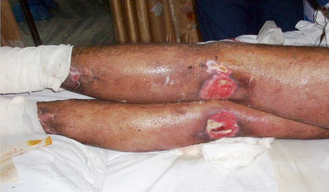
Figure 1: Bilateral popliteal fossa pressure sores.
On assessment, the pressure sores were classified as grade 2 (Wagner’s grading). His routine blood analysis showed elevated liver enzymes and severe metabolic acidosis. Multiple microorganisms (E. coli, K. pneumoniae, and Staph. heamolyticus) were cultured from the wounds. Skin biopsy from the edge of the sore showed ulceration of epithelium with underlying inflammation. No specific etiology was found. On evaluation, he was diagnosed with septic shock due to chest infection. He was intubated and kept in ICU on ventilatory and inotropic support for approximately one month, but his condition kept on deteriorating and he expired due to septicemia with multiple organ dysfunctions.
Discussion
Pressure ulcers are frequent complications of spinal cord injury. Pressure ulcer may develop anytime after spinal cord injury and are among the costliest complications. They are responsible for an increased length of hospital stay and are an important cause of rehospitalization.7 Disease or injuries of the spinal cord lead to knee, heel, or elbow spasticity in the lower or upper extremities, which can progress to increased muscle tone and pressure over bony prominence and subsequent pressure ulcer formation.8
Pressure ulcers typically occur over a bony prominence and the majority of ulcers are located in the low back area, including sacrum, coccyx, ischium, buttocks, greater trochanters and the leg area including the heels and lateral malleoli.4 These locations account for 87% of all pressure ulcers.5 Atypical pressure ulcers are generally not located over a bony prominence; they can be found in unusual places such as the nape of the neck and on the penis, nostrils, helix of the ears, or the upper back.9 Atypical pressure ulcers may result either due to the use of medical devices, increased spasticity (contracture), or bone deformity.9
As most paraplegic patients develop flexion contracture, pressure ulcers are usually seen over the extensor surface of the knee joint. Pressure ulcer over the popliteal fossa is very rare. Only one case has been reported in literature that was due to use of thromboembolic deterrent (TED) stockings.10 It is postulated that the combined effect of loss of sensation, diabetes, and septicemia- induced generalized body edema would have been responsible for pressure sores over the popliteal region.
Conclusion
Atypical pressure ulcers are generally not located over a bony prominence; they can be found in unusual places. Atypical pressure ulcers may result either due to the use of medical devices, increased spasticity (contracture) or bone deformity, so surgeon and nursing staff needs to be vigilant for pressure ulcers occurring at such unusual sites especially in paraplegic patients for early and appropriate search of etiological factors and their correction.
Acknowledgements
The authors reported no conflict of interest and no funding was received on this work.
|
|
| |
References
1. Allman RM, Goode PS, Patrick MM, Burst N, Bartolucci AA. Pressure ulcer risk factors among hospitalized patients with activity limitation. JAMA 1995 Mar;273(11):865-870.
2. Inouye SK, Studenski S, Tinetti ME, Kuchel GA. Geriatric syndromes: clinical, research, and policy implications of a core geriatric concept. J Am Geriatr Soc 2007 May;55(5):780-791.
3. Grey JE, Harding KG, Enoch S. Pressure ulcers. ABCs of wound healing. Non-surgical and drug treatments. BMJ 2006;332(7539):72-75.
4. Allman RM. Pressure ulcer prevalence, incidence, risk factors, and impact. Clin Geriatr Med 1997 Aug;13(3):421-436.
5. Smith DM. Pressure ulcers in the nursing home. Ann Intern Med 1995 Sep;123(6):433-442.
6. Jaul E. Assessment and management of pressure ulcers in the elderly: current strategies. Drugs Aging 2010 Apr;27(4):311-325.
7. Cardenas DD, Hoffman JM, Kirshblum S, McKinley W. Etiology and incidence of rehospitalization after traumatic spinal cord injury: a multicenter analysis. Arch Phys Med Rehabil 2004 Nov;85(11):1757-1763.
8. Knight DB, Scott H. Contracture and pressure necrosis. Ostomy Wound Manage 1990 Jan-Feb;26:60-62, 65-67.
9. Jaul E. A prospective pilot study of atypical pressure ulcer presentation in a skilled geriatric nursing unit. Ostomy Wound Manage 2011 Feb;57(2):49-54.
10. Ong JC, Chan FC, McCann J. Pressure ulcers of the popliteal fossae caused by thromboembolic deterrent stockings (TEDS). Ir J Med Sci 2011 Jun;180(2):601-602.
|
|