|
Distinguishing sarcoidosis from pulmonary tuberculosis can sometimes be a great challenge to physicians, especially in developing countries where there is high prevalence of tuberculosis. Both tuberculosis and sarcoidosis are granulomatous diseases, however, tuberculosis has a caseating granuloma as opposed to sarcoidosis, which present with non-caseating epithelioid cell granuloma. Due to the marked clinico-radiological similarity of these entities and high prevalence of tuberculosis, these patients receive repeated courses of anti-tubercular therapy (ATT) while lung damage continues to progress.
Recently, a 51 year old man presented to the out-patient department at Firoz Specialist Hospital, with one year history of non-productive cough, on and off low grade fever, and 9 kg weight loss. He was on ATT for the past one month. He had a past history of pulmonary tuberculosis 15 years before, for which he had a complete course of ATT. Respiratory system examination revealed inspiratory crept throughout in the right lung. Laboratory investigations showed normocytic normochromic anemia, white blood count 7530/mm3, erythrocyte sedimentation rate 45 mm at the end of one hour. Sputum for acid fast bacilli was negative.
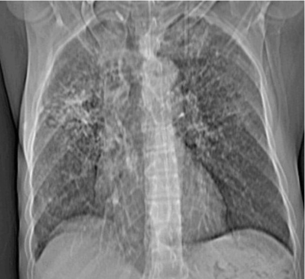
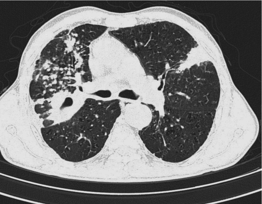
Chest radiography showed inhomogeneous opacity with reticulations in the right upper-mid zone (perihilar region), as shown in Fig. 1. High resolution Computed Tomography (HRCT) of the thorax revealed a cavitatory lesion in the posterior segment of the right upper lobe with multiple nodules in the anterior segment of the upper lobe and the middle lobe (Fig. 2). It was also associated with thickening of the interlobular septa and peri-bronchovascular bundle. Radiological differential diagnosis would be tuberculosis, sarcoidosis and lymphangitis carcinomatosis. However, secondary reactivation of tuberculosis was strongly suspected considering both clinical and radiological (mainly the cavitatory lesion) findings. Computed tomography guided lung biopsy revealed the presence of non-caseating granulomas suggestive of sarcoidosis. The patient was treated with prednisolone 60 mg/day. Within 3 weeks, marked clinical improvement was seen. Steroid was gradually tapered over the period of 4 weeks. The patient remained asymptomatic during the next six months.
Sarcoidosis is a multisystem disorder of unknown etiology, characterized by the presence of non-caseating granuloma.1 Non-specific constitutional symptoms such as fever, fatigue, malaise and weight loss are present in approximately one-third of patients. The chest radiograph usually shows hilar and mediastinal lymphadenopathy. Other characteristic findings include interstitial lung disease, or occasional calcification of affected lymph nodes. Most commonly, high resolution CT of the thorax shows thickening of bronchovascular bundles and perilymphatic distribution of nodules which represent the sarcoid granuloma. Clinical and radiological features of tuberculosis and sarcoidosis are quite overlapping and therefore, a diagnostic dilemma often persists. Exclusion of tuberculosis is important, particularly because corticosteroids form the mainstay of treatment for sarcoidosis. Cavity formation is common in tuberculosis and uncommon in sarcoidosis. Greiner et al. reported only 3% of cases with cavity formation.2
The diagnosis is established most securely when clinico-radiologic findings are supported by histologic evidence of widespread non-caseating granulomas.1 Recently, adding to the diagnostic difficulty, there are also several reports of coexistence of sarcoidosis and tuberculosis in the same patients.3

Figure 1: Chest X-ray (PA)-shows reticulonodular opacity in right perihilar region.

Figure 2: HRCT thorax- shows a thick walled cavitatory lesion with multiple nodules in right middle lobe along with thickening of interlobular septa and bronchovascular bundle. Also patch of consolidation seen in lingular lobe.
Acknowledgements
The author reported no conflict of interest and no funding was received for this work.
|