A Palatal Mass
Rashid Al-Abri,1 Sudesh Kumar,1 Salim Al-Sudairi2
Al-Abri R, et al. OMJ. 25, 55-56 (2010); doi:10.5001/omj.2010.16
From the 1Department of ENT, Sultan Qaboos University Hospital, Sultanate of Oman 2Department of Oral Health,
Sultan Qaboos University Hospital, Sultanate of Oman.
Received: 18 Sep 2009
Accepted: 02 Nov 2009
Address correspondence and reprint request to: Dr. Rashid Al-Abri, Department of ENT,
Sultan Qaboos University Hospital, Sultanate of Oman
E-mail: ralabri@hotmail.com
INTRODUCTION
A 19 year old female patient presented to the ENT department with complaints of slow growing, painless palatal mass for three months duration. On examination, there was a smooth surface 3x3 cm mass arising from the left side of hard palate (Fig. 1) which was firm in consistency on palpation. The rest of ENT examinations were normal.

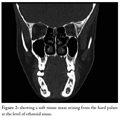
The Computed tomography (CT) showed a well defined contrast enhancing mass with no obvious bony erosion, (Fig. 2). The fine needle aspiration cytology from the lesion showed the histological features of epithelial and myoepithelial elements arranged in a variety of patterns and embedded in a mucopolysaccharide stroma.